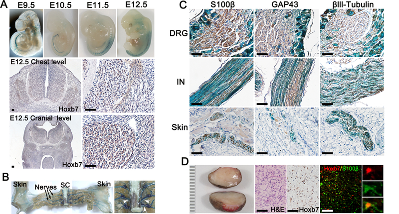
Figure 1. Hoxb7 lineage-derived cell populate nerve ending in the dermis.

(A-C) X-Gal staining was performed on E9.5 – E12.5 embryos (A) and adult mice (B-C) with genotype Hoxb7-Cre;LacZ. (A) Immunohistochemistry using anti-HOXB7 antibodies on transectional cut of E12.5 embryo at the chest (middle panels) and cranial (lower panels) level. (B) Gross dissection of adult mice peripheral nervous system. Black arrow indicates dorsal sensory nerves. White arrow heads indicates ventral roots. SC=Spinal Cord. (C) Immunohistochemistry using Schwann cell (S100β, GAP43) and neuronal (βIII tubulin) markers on histological section of dorsal root ganglion (DRG), intercostal nerve (IN) and skin X-Gal counterstained (blue). (D) Typical representation of human cNF (left panel). Scale bar in millimeters. Hematoxylin and eosin (H&E) and immunohistochemistry using anti-HOXB7 antibodies on human cNF (middle panels). Immunofluorescence on human cNF using anti-HOXB7 and anti-S100β antibodies. Scale bar = 50μm.
