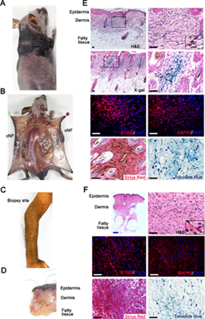
Figure 3. Ablation of Nf1 in Hoxb7 lineage cells give rise to diffuse cutaneous Neurofibroma.

(A) H7;Nf1mut mouse model bearing diffuse cNF. (B) Dissection of H7;Nf1mut mice showing that tumors are contained within the dermis. (C) NF1 patient with diffuse cNF of the forearm. (D) Biopsy of human diffuse cNF showing that tumor is contain within the dermis. (E-F) Histological characterization of murine (E) and human (F) diffuse cNF. Histochemistry was performed with X-Gal staining (Hoxb7 lineage tracing in mice), H&E, Sirius red (collagen), toluidine blue (mast cells), and immunofluorescence with anti-S100β and anti-GAP43 (Schwann cell markers). Black and white scale bar = 50μm. Blue scale bar = 1mm.
