
Fig. 2.
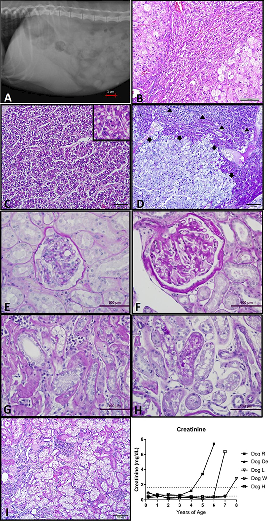
Liver and kidney disease in GSD Ia dogs. a. Hepatomegaly of Dog H noted on right lateral abdominal radiographs, liver far surpasses the border of the rib cage. b. Liver histology from Dog L with relatively normal parenchyma entrapped between two encroaching areas of focal nodular hyperplasia with marked hepatocyte enlargement and vacuolation (H&E 200x). c. Liver mass histology from dog De demonstrates nests and thickened trabeculae of neoplastic cells more prominent with absence of lobular architecture (H&E 200x). Inset box (400x) demonstrates a mitotic figure in center. d. Liver mass histology from dog L is composed of large, vacuolated neoplastic hepatocytes compresses and invades adjacent more normal hepatic parenchyma. Normal lobular architecture not present in neoplastic mass (adenoma, Arrows). There is a thin layer of compressed more normal hepatocytes above the adenoma. In upper most part of photomicrograph are nests and trabeculae thickened by neoplastic hepatocytes 2 or more cells wide (carcinoma, arrowheads). (PAS, 100x). e. Normal renal glomerulus (PAS, 200X). f. Glomerulus with changes of FSGS demonstrating thickened PAS positive glomerular basement membrane as well as thickened Bowman’s capsule (PAS 200x). g. Tubulo-interstitial changes as part of the spectrum of chronic renal lesions in dog H. Note interstitial mononuclear inflammatory infiltrate and thickened peritubular basement membrane. (PAS 200x) h. PAS positive droplets in proximal tubular epithelium (PAS 200x). i. Kidney histology from Dog R (100x) There is clustering of glomeruli with varying changes. In several glomeruli, there is moderate eosinophilic thickening of Bowman’s capsules (sclerosis). In other glomeruli, the sclerosis of the Bowman’s capsules is more severe and there is moderated to marked eosinophilic thickening of the mesangium of one or more segments of the glomeruli tufts. A sclerotic remnant of a glomerulus is also present. The proximal convoluted tubular epithelial cells are markedly swollen with clear, cytoplasmic vacuoles. j. Creatinine increased over time in dogs R, H and L.