
Fig. 6. Microtubule-dependent peripheral coupling supports endogenous BK channel activity and regulation of cerebral artery tone by intraluminal pressure.
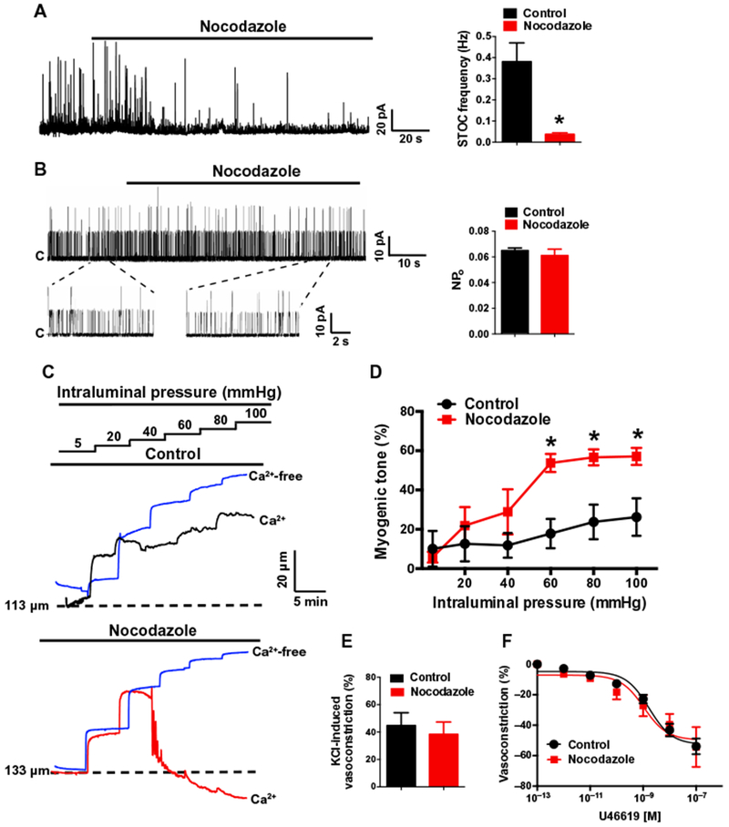
(A) Representative perforated voltage-clamp (VH = −30 mV) recording and summary data demonstrating the effects of nocodazole (10 μM) on STOC frequency (n = 5 cells, n = 3 animals; *P ≤ 0.05 compared to control). (B) Representative recordings and summary data demonstrating the effects of nocodazole (10 μM) on single BK channel activity. BK channel activity in inside-out membrane patches was evoked by 3 μM free Ca2+ in the bath solution and was recorded at +40 mV. Nocodazole had no effect on single-channel open probability (NPo) (n = 5 cells, n = 3 animals). (C) Representative recordings of pressure-induced constriction of control- and nocodazole-treated cerebral resistance arteries. Traces show inner luminal diameter at intraluminal pressures between 5 and 100 mmHg for arteries superfused with a Ca2+-containing (black or red traces) or Ca2+-free (blue trace) bathing solution. (D) Summary data showing the effects of nocodazole on myogenic tone as a function of intraluminal pressure (n = 5 arteries, n = 3 animals; *P ≤ 0.05 compared to control). (E) Summary of constriction of control- or nocodazole-treated arteries in response to increased (60 mM) extracellular [K+] (n = 4 arteries, n = 3 animals). (F) Summary concentration-response curves demonstrating that nocodazole treatment does not alter sensitivity to the vasoconstricting thromboxane receptor agonist U46619 (n = 4 arteries, n = 3 animals).