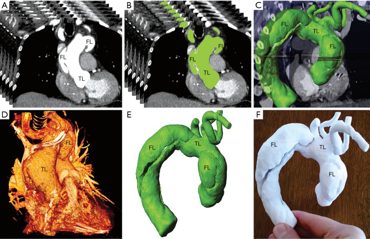
Figure 1.
Three-dimensional (3D) reconstruction of the aortic dissection of a patient, where the aortic tear located in all segments of the aorta with flap propagated to all segments of the aorta. (A) Computed tomography scan of the chest showing complete dissected aorta; (B) thresholding of the dissected aorta; (C) integration of segmented aorta with 3D CT image; (D) 3D surface rendering of the aorta and surrounding structures; (E) 3D segmentation of the aorta; (F) 3D-printed model of the TAAD. TAAD, type A acute aortic dissection; TL, true lumen; FL, false lumen. Reprinted with permission from Hossien et al. (35).

