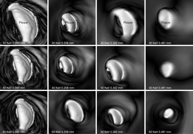
Figure 3.
3D virtual intravascular endoscopy images of plaque at left circumflex acquired with different beam energies and slice thicknesses. The plaque became irregular when the slice thickness of 0.491 mm was used for image reconstruction. Arrows indicate simulated thrombus in the main pulmonary arteries. Reprinted with permission from Sun et al. (38).

