Fig. 7.
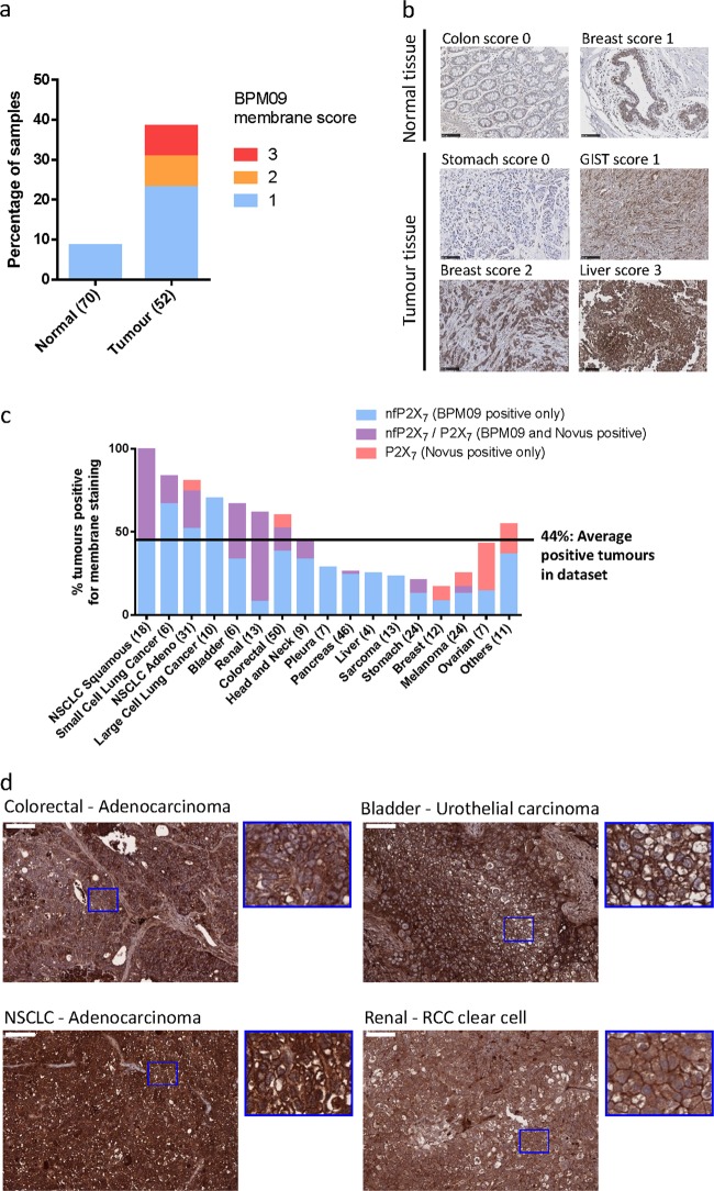
nfP2X7 is present in a broad range of tumour samples. a Tissue sections from 70 normal biopsies and 52 tumour biopsies were stained with BPM09 and scored for membrane staining. b Representative images for each score of normal and tumour tissue section stained with BPM09. Scale bar represents 100 µM. c Tissue sections from 290 Patient-derived xenograft (PDX) models were stained with BPM09 and a polyclonal antibody raised against part of the extracellular domain of P2X7a. Tissue sections were scored for membrane staining. For each tumour type the percentage of PDX with positive staining is shown with the number of PDX examined in brackets. d Representative images of colorectal (adenocarcinoma), bladder (urothelial carcinoma), non-small cell lung cancer (NSCLC—adenocarcinoma) and renal cancer (RCC clear cell) tissue sections with positive BPM09 staining together with higher magnification section showing BPM09 membrane staining. Scale bar represents 100 µm

