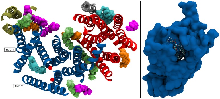
Figure 13.
(Left panel) TMDs of γ-secretase with strongly immobilized lipids. The TMDs of PS-1 are colored in dark blue, PEN-2 is depicted in beige, APH-1 in red and nicastin in gray. The lipids are colored according to their B-factor: Cyan: 156 to 250Å2; orange: 405 to 519Å2; green: 610 to 732Å2; purple: 785 to 1026Å2. The active site residues D257 and D385 are marked by red dots. (Right panel) Van der Waals surface depiction of presenilin-1 with the strongly bound POPC molecule in the cavity.

