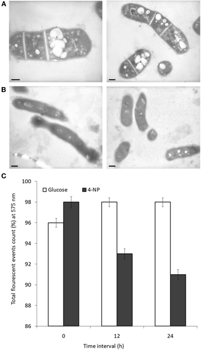
Figure 4.

Lipid inclusions observed during growth on glucose are lost during growth on 4-NP. TEM images of cells grown for 12 h on (A) glucose, or (B) 4-NP. Bar = 200 nm. (C) Flow cytometric quantification of lipid content of cells pre-grown on glucose, stained with Nile red (total event counts % for emission at 575 nm) at 0, 12, and 24 h after shifting to growth on 4-NP.
