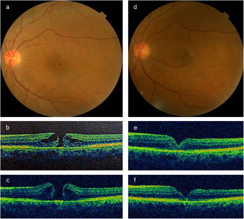
Fig. 3.
Fundus photographs and optical coherence tomography (OCT) images of the small full-thickness macular hole (FTMH) in left eye with a diameter of 191 μm, without vitreomacular traction (VMT), before (a, b) and 1 month (c) after intravitreal injection of expansile gas. The hole progressed to a medium FTMH with a diameter of 383 μm. After 1 month of intravitreal gas injection, the patient was treated by subsequent vitrectomy. An anatomical closure of the hole was demonstrated by fundus photograph and OCT images at 2 weeks (e) and 2 months (d, f). Corrected visual acuity was decreased from 0.3 (pre-treatment) to 0.1 (post-treatment)

