Figure 3.
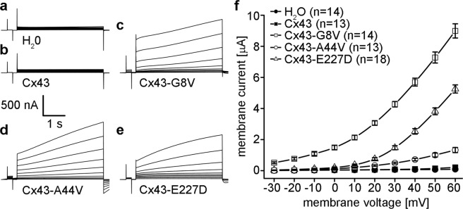
Cx43 mutations induce large hemichannel currents in Xenopus oocytes. Single cells were clamped at a holding potential of −40 mV and subjected to voltage pulses ranging from −30 to +60 mV in 10 mV steps. H2O (a) and wild-type Cx43 (b) injected cells displayed negligible membrane currents. Cx43-G8V (c), Cx43-A44V (d), and Cx43-E227D (e) expressing oocytes displayed much larger hemichannel currents than wild-type Cx43. (f) Steady-state currents from each pulse were plotted as a function of membrane voltage. Steady state currents in wild-type Cx43 (filled squares) or H2O injected control cells (filled circles) were negligible at all tested membrane voltages. Cx43-G8V (open squares) expressing cells exhibited significantly increased steady-state currents at all voltages compared to either control or wild-type Cx43 oocytes. Cx43-A44V (open circles), or Cx43-E227D (open triangles) currents were similar to those observed in control cells at negative voltages, but increased at positive potentials. Data are the mean ± SEM.

