Fig. 1.
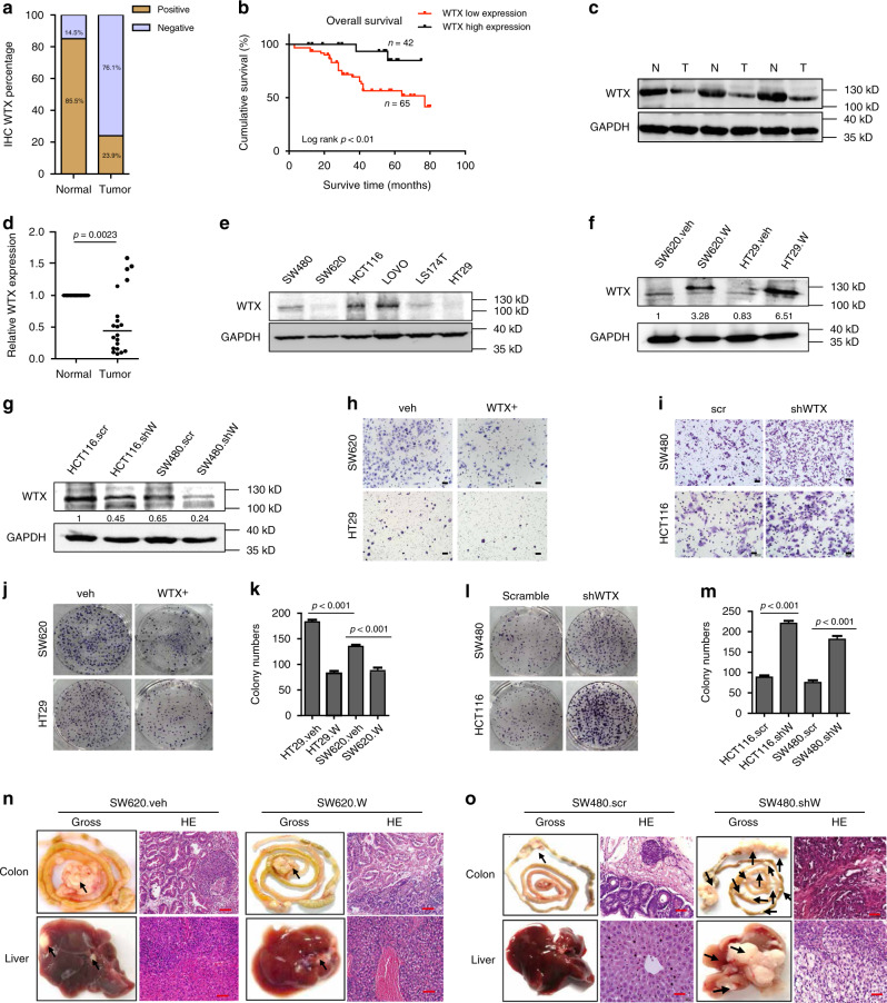
WTX loss promotes CRC cells, migration and liver metastasis. a Percentage of WTX IHC in CRC and matched adjacent normal colorectal mucosa tissues. b Kaplan–Meier survival analysis of CRC patients with WTX high or WTX low expression. c Imunoblotting (IB) analyzes the WTX expression in human CRC tumor (T) and matched adjacent normal mucosa (N) tissues. d qRT-PCR analyzes WTX mRNA level in human CRC tumor and matched adjacent normal mucosa tissues. **p = 0.0023, mean ± SEM. Two-tailed Student’s t-test. e IB analyzes WTX expression in CRC cell lines. f, g IB analyzes WTX expression in indicated cell lines. h, i Transwell analyzes the migration of the indicated cell lines. Scale bars, 200 μm. j–m Colony formation analysis of the indicated cell lines. ***p < 0.001, mean ± SEM. Two-tailed Student’s t-test. n, o Gross and H&E staining observe the CRC orthotopic tumors and liver metastasis in indicated cell lines. Each arrow pointed a tumor mass. Scale bars, 20 μm, n = 11 (n) and n = 8 (o)

