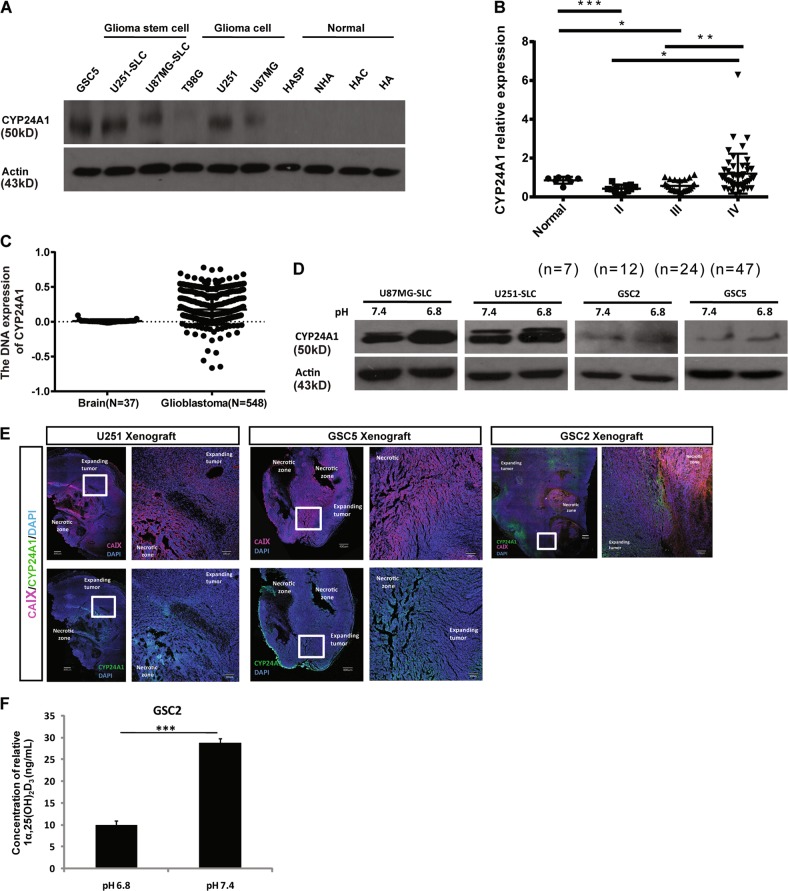
Fig. 4. CYP24A1 was highly expressed in acidic microenvironment and high grade glioma tissues.
a Immunoblotting of the expression of CYP24A1 in HA, HAC, NHA, and HASP normal human astrocytes, U87MG, U251, and T98G glioma cell lines and U87MG-SLC, U251-SLC, and GSC5 stem cell-like glioma cells. b The relative CYP24A1 protein levels in seven normal brain tissues and in 83 glioma tissues (12 grade II, 24 grade III, and 47 grade IV glioma tissues); actin was used as a control. The relative intensity values of CYP24A1/actin were analyzed by Image J software. c Immunoblotting of the expression of CYP24A1 in U87MG-SLC, U251-SLC, GSC2, and GSC5 cells under pH 7.4 or pH 6.8 conditions. d Immunofluorescence analysis of CYP24A1 (green) and carbonic anhydrase IX (CA IX, red) merged with nuclear DAPI staining (blue) in xenografts developed from U251, GSC2, and GSC5 cells (bar = 400 μm, left; bar = 100 μm, right). e Quantification of endogenous 1α,25(OH)2D3 in pH 7.4-treated and pH 6.8-treated GSC2 cells. The values shown are the means ± SD of at least three independent experiments (***P < 0.001, Student’s t-test)

