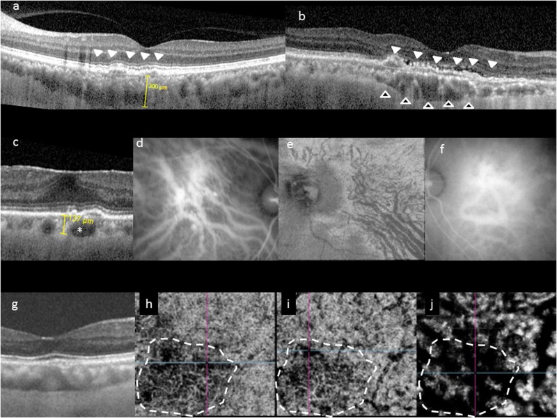
Fig. 1.
Choroidal features common to pachychoroid phenotype. Typical choroidal features in the pachychoroid disease phenotype include diffuse or focal increase in choroidal thickness, which is typically associated with abnormally dilated Haller layer vessels (pachyvessels) and attenuation of the inner choroid (Sattler’s layer and choriocapillaris). Choroidal thickness can be readily evaluated in cross-sectional optical coherence tomography (OCT), which may show diffuse thickening and increased subfoveal choroidal thickness (a), or focal thickening (b, hollow arrowheads). In some eyes, an irregular elevation of the retinal pigment epithelium (RPE) can be seen to overlie these choroidal abnormalities (white arrowheads). Pachyvessels can be identified as a choroidal vessel with enlarged caliber (c, *) which can occupy almost the entire thickness of the choroid. Pachyvessels can also be seen as dilated submacular vessels which do not taper toward the posterior pole on ICGA (d) or on en face OCT (e). These pachyvessels may be distributed in a diffuse (d) or patchy manner (e). Pachyvessels usually exhibit choroidal vascular hyperpermeability with indocyanine green angiography (ICGA) (f) which may suggest choriocapillaris ischemia. Using OCT angiography (OCTA), the spatial correlation between choriocapillaris blood flow and pachyvessels can be evaluated in a depth-resolved manner. In g–j, choroidal thickening and choriocapillaris attenuation seen in cross-sectional OCT (g) can be correlated with OCTA findings showing attenuation of flow signal (dash white outline) within the choriocapillaris (h) and inner choroid (i) which directly overlie areas with dilated outer choroidal vessels (j)

