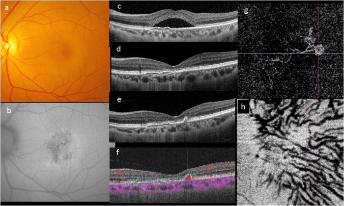
Fig. 3.
Evolution of a case over 4 years. A 46-year-old woman first presented with a solitary neurosensory detachment and mottled fundus autofluorescence (a–c). A shallow elevation of the RPE was noted. The SRF resolved spontaneously within 3 months and the eye remained unchanged for the following 4 years (d). During routine follow-up, development of a narrow-peaked PED without SRF was noted on OCT (e). OCTA showed a localized area of abnormal flow signal within the narrow-peaked PED (f). En face OCTA showed a neovascular network with aneurysms its temporal margin (g). The outline of pachyvessels can be seen as dark silhouettes in the OCTA segmented through Haller’s layer (h)

