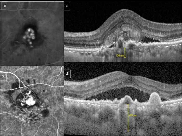
Fig. 4.
Choroidal features in polypoidal choroidal vasculopathy (PCV)/aneurysmal type 1 neovascularization (AT1). A bimodal distribution of choroidal thickness has been described in PCV/AT1. This figure shows two eyes with PCV/AT1 confirmed with indocyanine green angiography (ICGA) (a, b). Subfoveal choroidal thickness was 130 and 420 μm, respectively (c, d). However, in both cases, choriocapillaris/inner choroid (outline by dashed white line in preserved areas) appears to be compressed and attenuated by underlying outer choroidal vessels in the subfoveal area (c, d)

