Fig. 3.
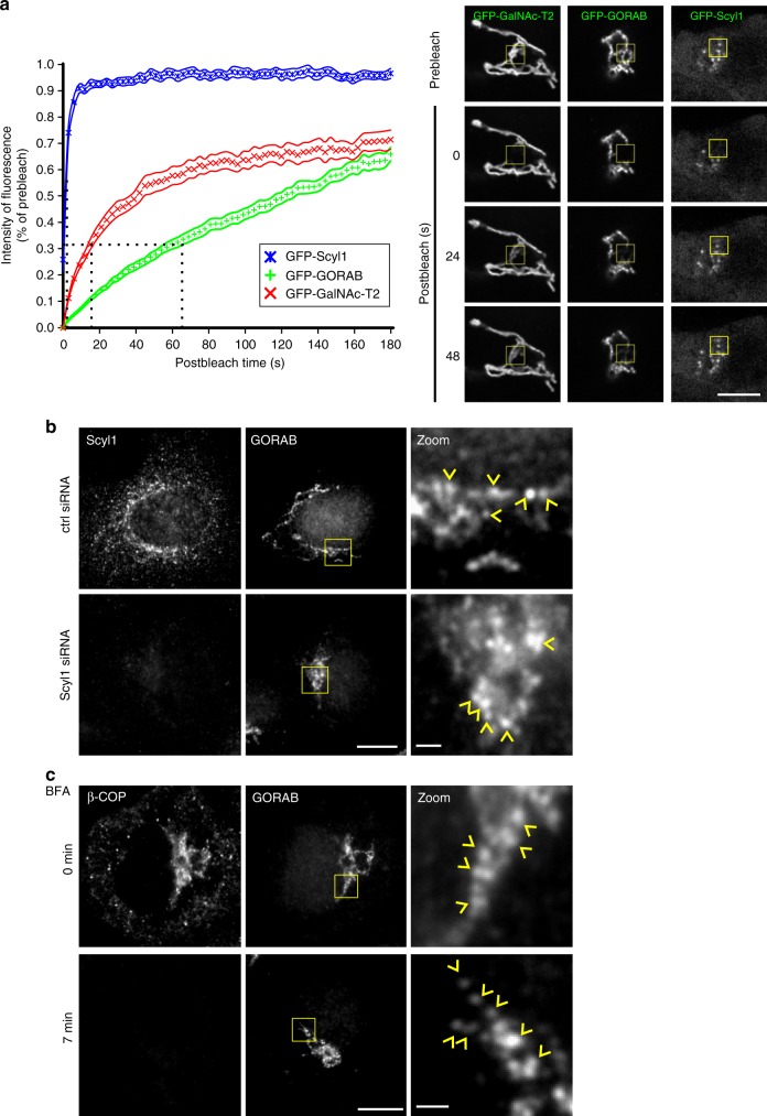
GORAB domains are stable entities. a Fluorescence recovery after photobleaching. Left, FRAP recovery curves for GFP-GalNAc-T2, GFP-GORAB and GFP-Scy1. Means with SEM for GFP-GalNAc-T2 (n = 27 cells), GFP-GORAB (n = 28 cells) and GFP-Scyl1 (n = 18 cells). Dotted lines mark points of half-time recoveries. Right, representative HeLa GFP-GalNAc-T2, HeLaM GFP-GORAB and HeLaM GFP-Scyl1 cells at pre-bleached and selected post-bleached states. Bleached region of interests are marked with yellow boxes. Scale bar, 10 µm. b Localization of GORAB in Scyl1-depleted cells. HeLa cells transfected with control or Scyl1 siRNA were fixed and labeled with antibodies to Scyl1 and GORAB. Scale bars, 10 µm and 1 µm. GORAB domains are marked with yellow arrowheads. c Localization of GORAB in BFA-treated cells. HeLa cells were exposed to 5 µg/mL BFA for 7 min prior to fixation and labeling with antibodies to Scyl1 and GORAB. Scale bars, 10 µm and 1 µm. GORAB domains are marked with yellow arrowheads

