Fig. 6.
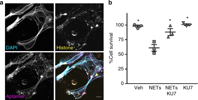
Aptamers bind to human neutrophil-derived NETs and inhibit NET-induced cytotoxicity. a Confocal microscopy of human neutrophil-derived NETs. Single images are shown in gray scale. DAPI (top, left); histones (top, right); aptamer KU7 (bottom, left); merged images show: cyan, DAPI labeling of DNA, yellow, histones, and magenta, aptamer KU7–647 (bottom, right). White areas in the merged image represent close proximity of DNA, histone and aptamer. Representative images, captured with 40× oil and 2.8× zoom. Scale is equivalent to 10 µm. b Aptamer inhibition of NETs-mediated cytotoxicity of endothelial cells determined by MTS assay. EA.hy926 cells treated with 8 µg per well of NETs material (based on DNA concentration) and/or 8 µg per well (10.66 µM) of aptamer (KU7); *p < 0.01 vs. NETs, one-way ANOVA corrected for multiple comparisons (Tukey), n = 3, data represent mean ± SEM

