Fig. 5.
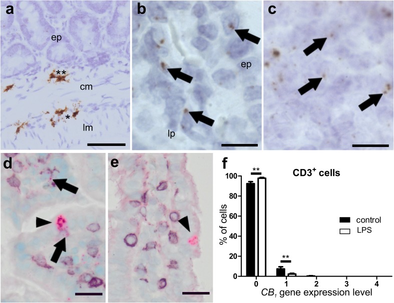
ISH staining of CB1 mRNA in ileum of healthy (a–d) and LPS-treated (e) mice. Strong CB1 mRNA ISH staining (brown, DAB) is seen in myenteric (asterisk) and submucosal ganglia (double asterisk) in the ileal wall of healthy mice (a). There is only low expression of CB1 mRNA (arrows) in the epithelium (bep) and the lamina propria (blp). High magnification of lymph follicles shows CB1 gene expression in healthy animals (c). High levels of CB1 mRNA (FastRed) are detected in isolated cells of the epithelium, probably neuroendocrine cells (arrowheads), from healthy (d) as well as LPS-treated (e) mice. Only marginal staining of CB1 mRNA is found in CD3+ cells (d, purple, VIP; arrows) that is even less in the ileum of LPS-treated animals (e). Quantification of CB1/CD3+ double staining is shown in f. Calibration bar in a: 50 µm; calibration bar in b, c: 10 µm; calibration bar in d, e: 20 µm; cm circular muscle layer, ep epithelium, lp lamina propria, lm longitudinal muscle layer; *, myenteric ganglion; **, submucosal ganglion

