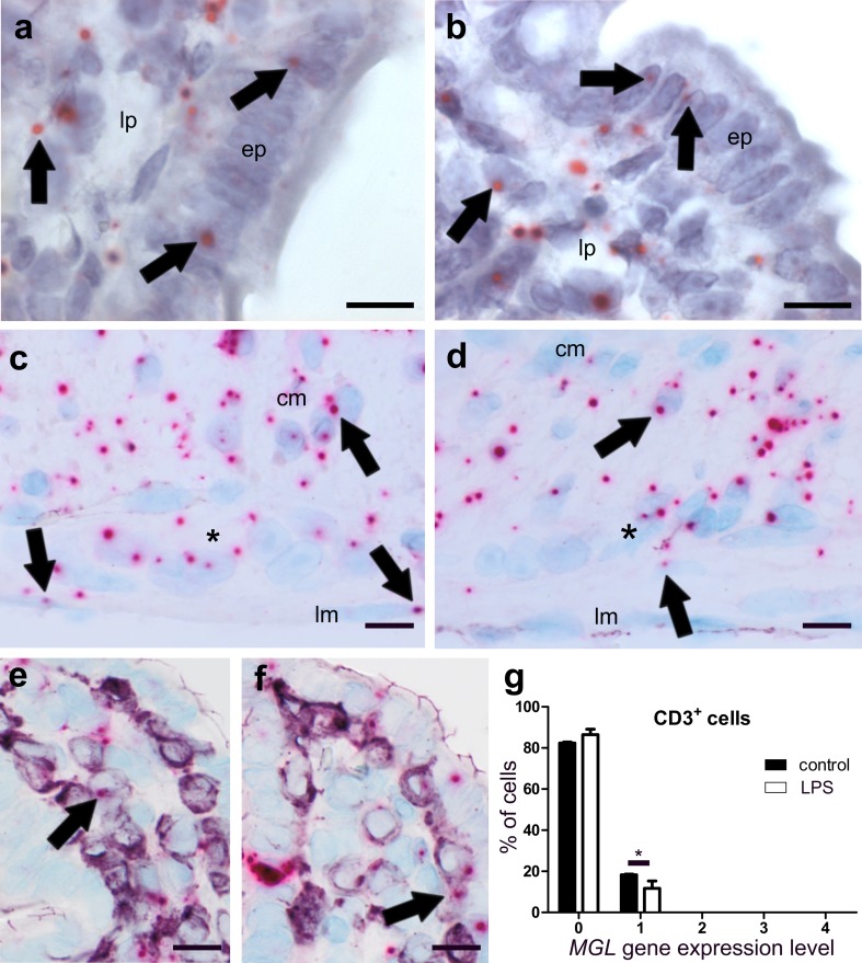
Fig. 7.
ISH staining of MGL mRNA (FastRed) in ileum of healthy (a, c, e) and LPS-treated (b, d, f) mice. MGL mRNA is equally present in epithelium (ep) and lamina propria (lp) cells of healthy ileum (a) and ileum from LPS-treated mice (b arrows denote representative mRNA staining). Expression is also present in myenteric ganglia (denoted by an asterisk) of healthy (c) and LPS-treated (d) mice. Expression of the MGL gene is widespread in the circular (cm) and the longitudinal muscle layer (lm), (arrows in c), and it is slightly decreased in LPS-treated mice (d). Only low co-localization of MGL gene expression with CD3+ cells (purple, VIP) is found in healthy (e) and even lower co-localization in LPS-treated (f) mice. Quantification of MGL/CD3 double staining is shown in g. Calibration bars: 10 µm; cm circular muscle layer, ep epithelium, lp lamina propria, lm longitudinal muscle layer, cl crypt lumen; *, myenteric ganglion

