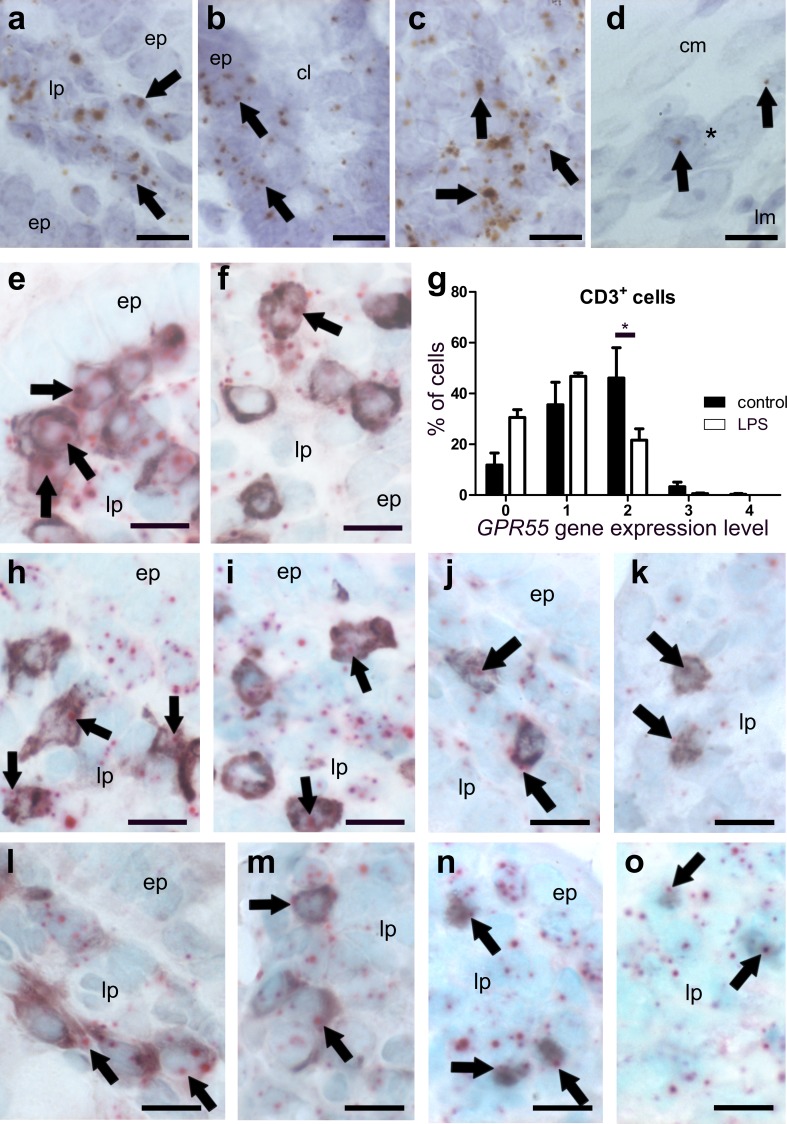
Fig. 8.
ISH staining of GPR55 mRNA (brown, DAB, a–d) and co-localization of GPR55 mRNA (FastRed, e–o) with markers for T-lymphocytes and macrophages (all purple, VIP) in the ileum of healthy (a–d, e, h, j, l, n) and LPS-treated mice (f, i, k, m, o). GPR55 mRNA is predominantly found in the lamina propria of the ileum mucosa (arrows point at representative expression) and in epithelial cells of healthy mice (a, b). Epithelial cells (ep) of villi show low levels of GPR55 mRNA (a), whereas crypt epithelial cells (ep) display relatively strong GPR55 gene expression (b). In healthy animals, strong GPR55 gene expression is detected also in lymph follicles (c), while in myenteric ganglia, expression is low (d; arrows; ganglion denoted by asterisk). GPR55 gene expression appears to be stronger in CD3+ T-cells of the lamina propria (lp) of the healthy ileum (e) when compared to LPS-treated animals (f). Arrows denote representative staining. Quantification of GPR55 mRNA in CD3+ cells is shown in g. In the lamina propria of the ileum mucosa, GPR55 gene expression is seen in CD4+ cells (h, healthy; i, LPS-treated), CD8+ cells (j, healthy; k, LPS-treated), F4/80+ cells (l, healthy; m, LPS-treated), and FoxP3+ T regulatory cells (n, healthy; o, LPS-treated). Calibration bars: 10 µm; ep epithelium, lp lamina propria, *, myenteric ganglion; arrows point at representative cells expressing the GPR55 gene

