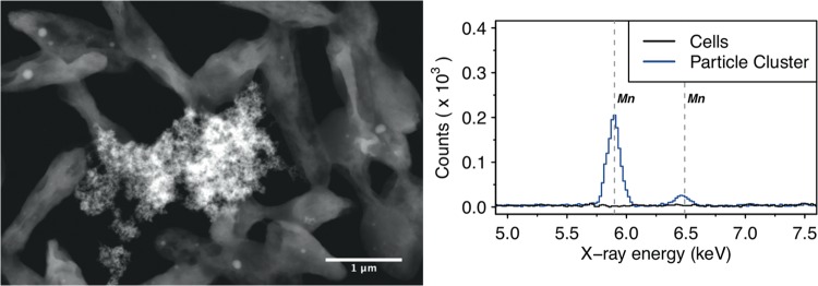
FIG 6.
Scanning transmission electron micrograph (left figure, high-angle annular dark field) of (granular) manganese-containing precipitate (center) surrounded by AzwK-3b cells, and associated energy-dispersive X-ray spectroscopic analysis (right figure) in this location. Only the energy range containing the manganese-specific X-ray energies at 5.90 keV (KαI) and 6.49 keV (KβI) is shown, and the manganese transitions are indicated by vertical gray dashed lines.

