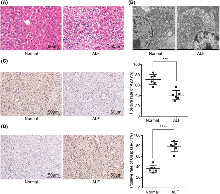
Figure 2. HE staining, ultrastructure observation, and immunohistochemistry show that ALF rats display serious pathological injury, lower positive expression rate of A20 protein, and higher that of Caspase-3 protein.
(A) Image of liver tissues in normal and ALF rats using HE staining (×200); (B) ultrastructure observation of hepatocytes in normal and ALF rats (×20000); (C) positive expression rate of A20 protein in normal and ALF rats using immunohistochemistry (×200); (D) positive expression rate of Caspase-3 protein in normal and ALF rats using immunohistochemistry (×200); measurement data were expressed as mean ± standard deviation; the data were assessed by t-test; n=6. ***, P<0.001; ****, P<0.0001.

