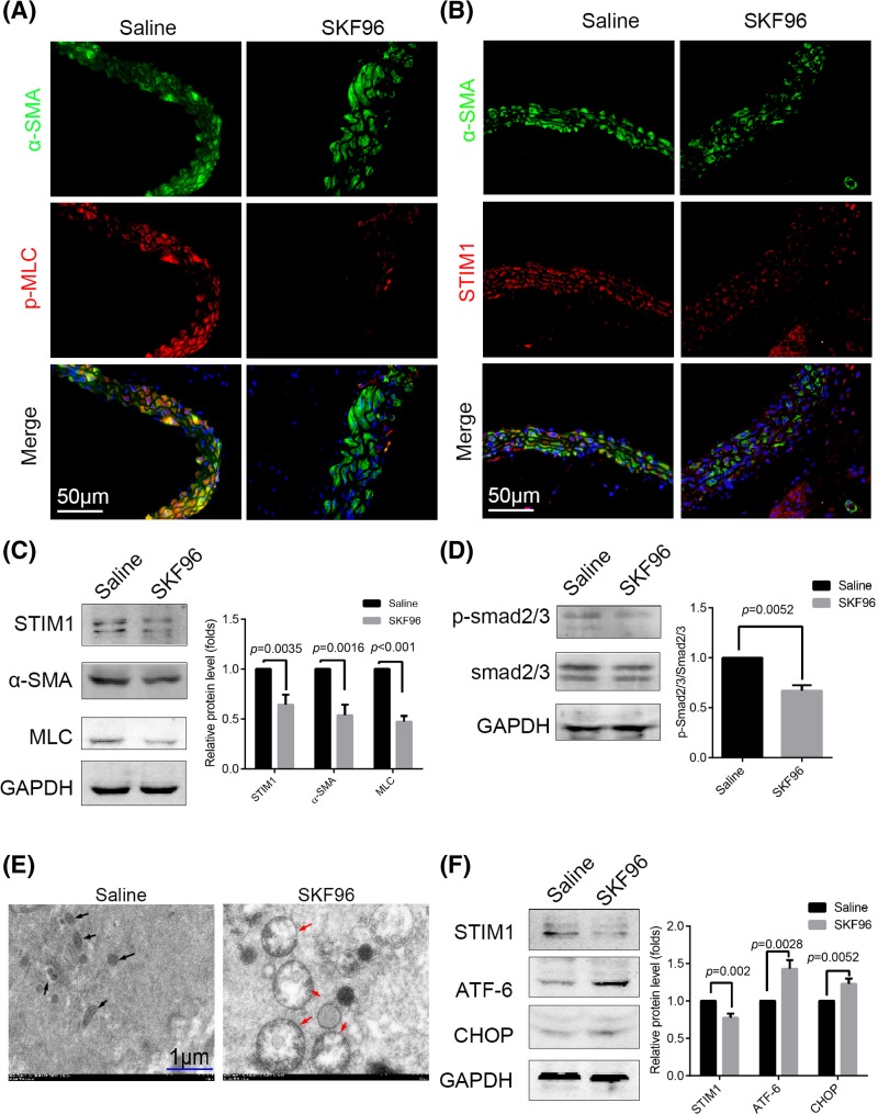
Figure 1. SKF96365 reduced STIM1 expression, impaired mitochondria, and led to ER stress in ASMCs in vivo.
ApoE−/− mice were treated with SKF96365 (n=6) or saline (n=6) by intraperitoneal injection for 1 week. (A,B) Immunofluorescence images of p-MLC and STIM1 in ASMCs (marked by α-SMA staining) after SKF96365 treatment for 1 week; n=6. (C) Presence of STIM1, α-SMA, and MLC as determined in SKF96 and control group aortic samples. (D) Phosphorylation of smad2/3 and expression of total smad2/3 assessed by Western blotting in the two groups. (E) ASMCs of the SKF96 group exhibited apparent swelling and deformation of the mitochondria (red arrows) compared with the saline group (black arrows), as observed by TEM. (F) Expression of the ER stress-related proteins ATF-6 and CHOP was elevated in the aortic samples of SKF96365-treated mice by Western blotting. All Western blotting analyses were performed three times. Representative images are shown. Data are represented as mean ± S.D.(t test, two-sided).

