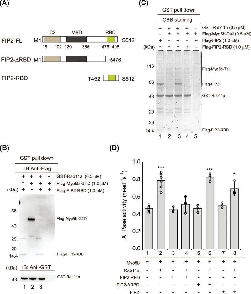
Figure 4. FIP2 bridges Myo5b and Rab11a.
(A) Schematic FIP2 constructs; C2, phospholipid-binding C2 domain; MBD, Myo5-binding domain; RBD, Rab11a-binding domain. (B) FIP2–RBD competes with Myo5b–GTD in binding with Rab11a. The interaction between GST–Rab11a and Flag-Myo5b-GTD in the absence or presence of Flag-FIP2-RBD was analyzed by GST pull down assays. The pulled down samples were analyzed by Western blot using Anti-Flag and Anti-GST antibodies. (C) FIP2 enhances the interaction between Rab11a–FIP2 and Myo5b–tail. GST pull down of GST–Rab11a with Flag-Myo5b-tail and Flag-FIP2 constructs. The GST pulled down samples were separated by SDS–PAGE and visualized by Coomassie Brilliant Blue Staining. (D) Effects of Rab11a and different FIP2 constructs on Myo5b ATPase activity. The ATPase activity of Myo5b was measured as described in Figure 1D, except in the presence of 12 μM indicated proteins (GST–Rab11a and FIP2 proteins). Values are mean ± S.D. from at least three independent assays. * (P<0.05) and *** (P<0.001) indicate significant differences compared with the first column. The pull down assays (Figure 4B,C) were repeated three times and similar results were obtained.

