
Figure 1.
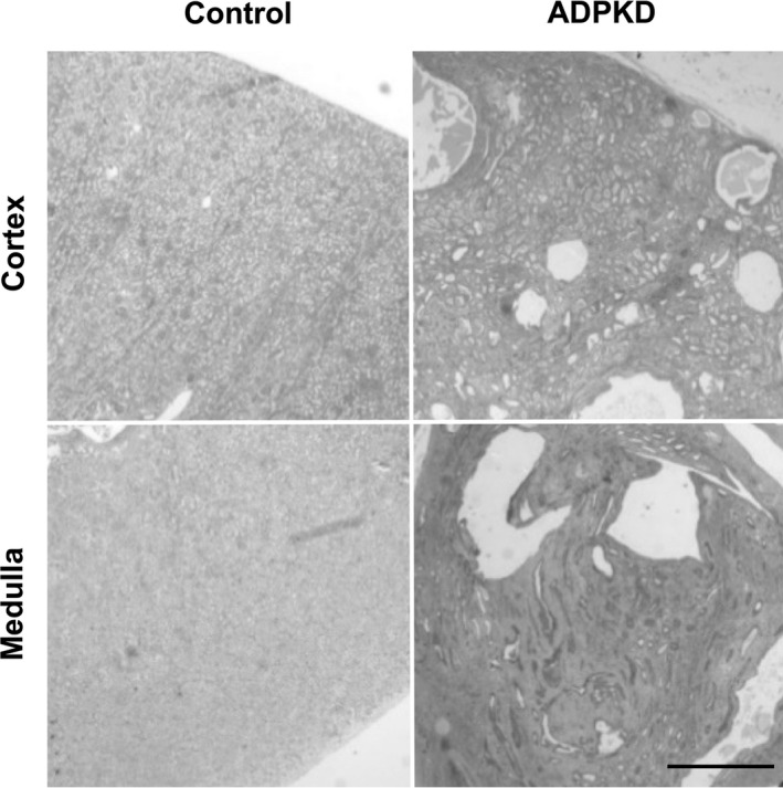
Examples of ADPKD and control renal tissues. Representative hematoxylin and eosin‐stained sections of tissues that were used in our study. These tissues contain relatively small amount of microscopic cysts and fibrosis because they were collected from normal‐appearing compartments of ADPKD kidneys. Scale bar = 1 cm.