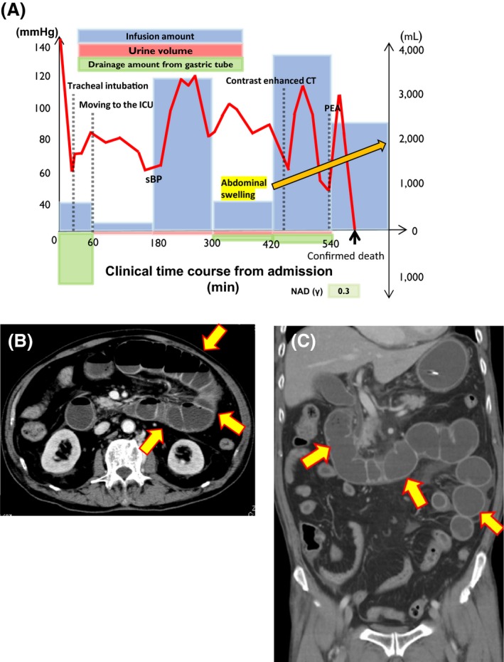
Figure 1.

Time course and imaging findings of a 79‐year‐old man with fulminant pseudomembranous enterocolitis caused by Klebsiella oxytoca. A, Schema of the patient's clinical time course (min) from admission to death. ICU, intensive care unit; NAD (γ), nor‐adrenaline (mg/kg/min); PEA, pulseless electrical activity; sBP, systolic blood pressure. B,C, Contrast‐enhanced computed tomography scan (6 h after admission). Arrows indicate intestinal dilatation and swelling, with the fluid level between the duodenum and the jejunum. However, there were no signs of ischemia or necrosis because the intestinal wall was well‐enhanced with contrast media.
