
Figure 4. Increased p62 immunolabeling in striatal ChIs.
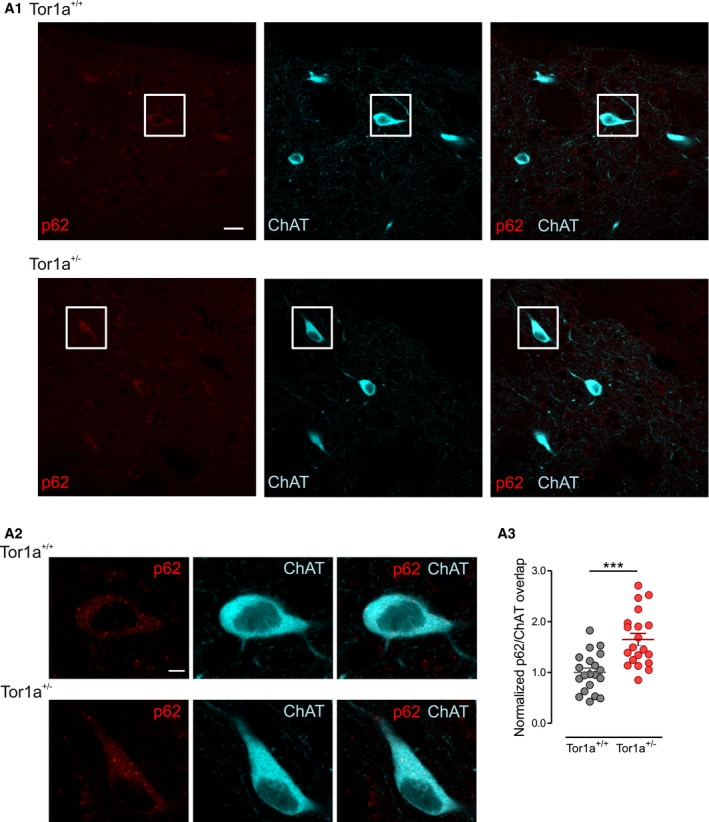
Representative double‐immunofluorescence confocal images of coronal sections of the dorsal striatum showing p62 immunolabeling (red) in choline acetyltransferase (ChAT)‐positive (cyano) ChIs of Tor1a +/+ and Tor1a +/− mice. (A1) Two representative ChIs identified by the white box in the low‐magnification images (scale bar = 20 μm) are shown at higher magnification in (A2) (scale bar = 5 μm). (A3) The graph reporting mean ± SEM values of p62/ChAT signal overlap in ChIs, normalized to Tor1a +/+ values of the same experiment, shows a significant increase in p62 in Tor1a +/− neurons (Tor1a +/+ n = 20 cells, N = 6 mice; Tor1a +/− n = 20 cells, N = 6 mice, t‐test ***P = 0.00008).