Figure 7.
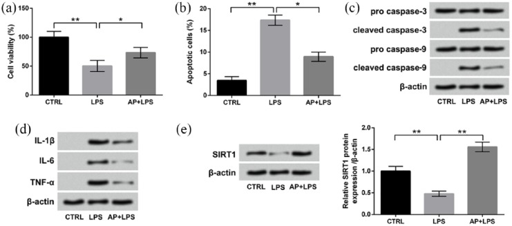
AP post-treatment also attenuated the LPS-induced HaCaT cell inflammatory injury and upregulated the expression of SIRT1. HaCaT cells were stimulated with 10 μg/mL LPS for 12 h and then exposed to 100 μg/mL AP for 24 h. Non-treated cells acted as control. Then, (a) cell viability, (b) percentage of apoptotic cells, (c) expression of proteins associated with apoptosis, (d) expression of inflammatory cytokines, and (e) expression of SIRT1 were measured by CCK-8 assay, flow cytometry assay, and western blotting, respectively. Data were presented as the mean ± SEM of three independent experiments. CTRL: control.
*P < 0.05; **P < 0.01.

