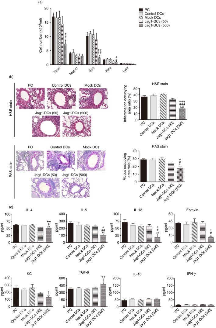
Figure 5.

Effect of Jag1‐dendritic cells (DCs) on airway inflammation and production of mediators in bronchoalveolar lavage fluid (BALF). (a) Each group of mice was killed after measuring pulmonary function parameters. Cells from the BALF were stained with Liu's dye. Numbers of total cells, eosinophils, neutrophils, macrophages, and lymphocytes were counted. (b) Lung sections were stained with H&E and PAS to measure inflammatory cells and mucus production around the airway. Inflammatory changes and mucus production are respectively presented as percentages of the inflamed area and the PAS‐positive area. (c) BALF was collected and analyzed for interleukin‐4 (IL‐4), IL‐5, IL‐10, IL‐13, interferon‐γ (IFN‐γ), transforming growth factor‐β (TGF‐β), eotaxin, and keratinocyte‐derived chemokine (KC) contents by ELISA kits. Results are expressed as the mean ± SEM of five mice in each group. *P < 0·05, **P < 0·01, ***P < 0·001 versus the positive control (PC) group. # P < 0·05, ## P < 0·01, ### P < 0·001 versus the control DC group. + P < 0·05, ++ P < 0·01, +++ P < 0·001 versus the mock DC group.
