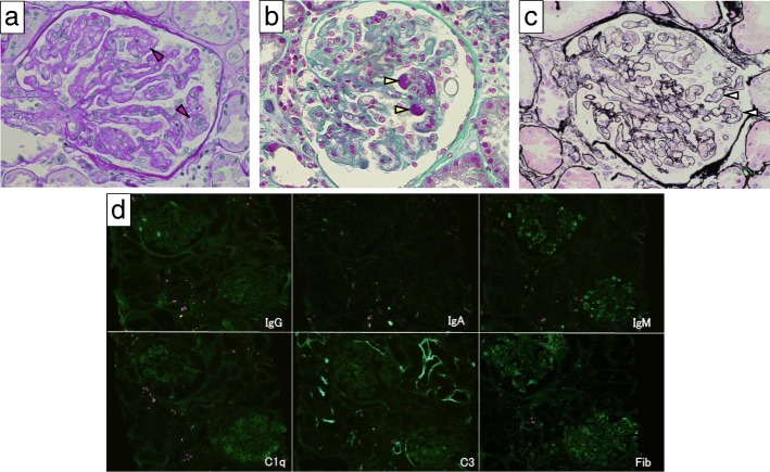
Fig. 2.
Renal biopsy findings of the case. a Periodic acid–Schiff staining showing mesangiolysis (red arrowhead). Original magnification 40X. b Azan staining showing fibrin thrombi (yellow arrowhead). 40X. c Periodic acid-methenamine silver staining showing double contour of the basement membrane (white arrowhead). 40X. d Immunofluorescence for IgG, IgA, IgM, C1q, C3 and fibrinogen (Fib). 20X

