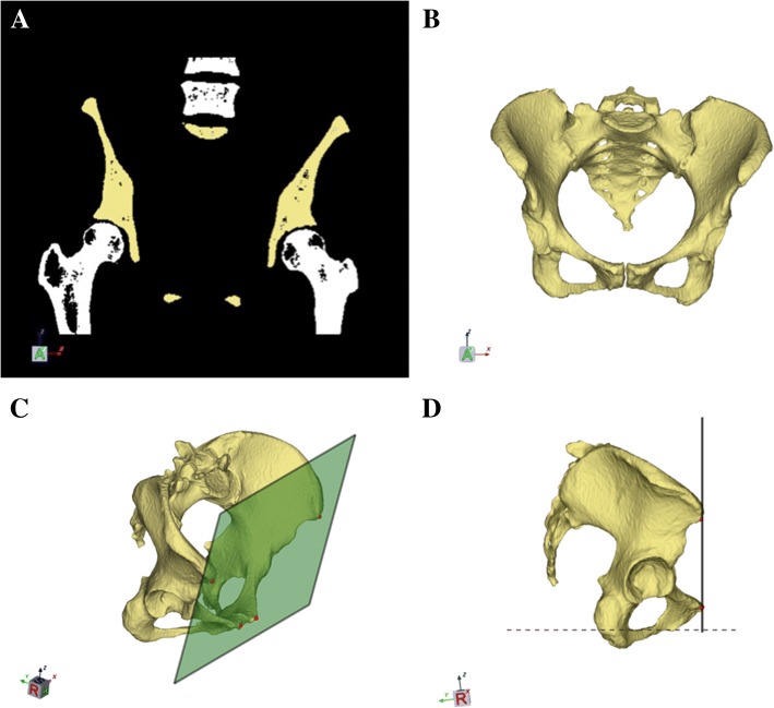
Fig. 1.
a–d Segmentation, modeling, and alignment of the 3D pelvic model. a A spherical mask was fitted to isolate the pelvis from CT volume images. Femurs and the lumbar vertebra were manually removed. b A virtual 3D pelvic model was automatically reconstructed using a threshold and region-growing algorithm. c Both anterior superior iliac spines and pubic tubercles were manually selected to determine the anterior pelvic plane. d The pelvic model with the anterior pelvic plane perpendicular to the horizontal plane

