
Fig. 6.
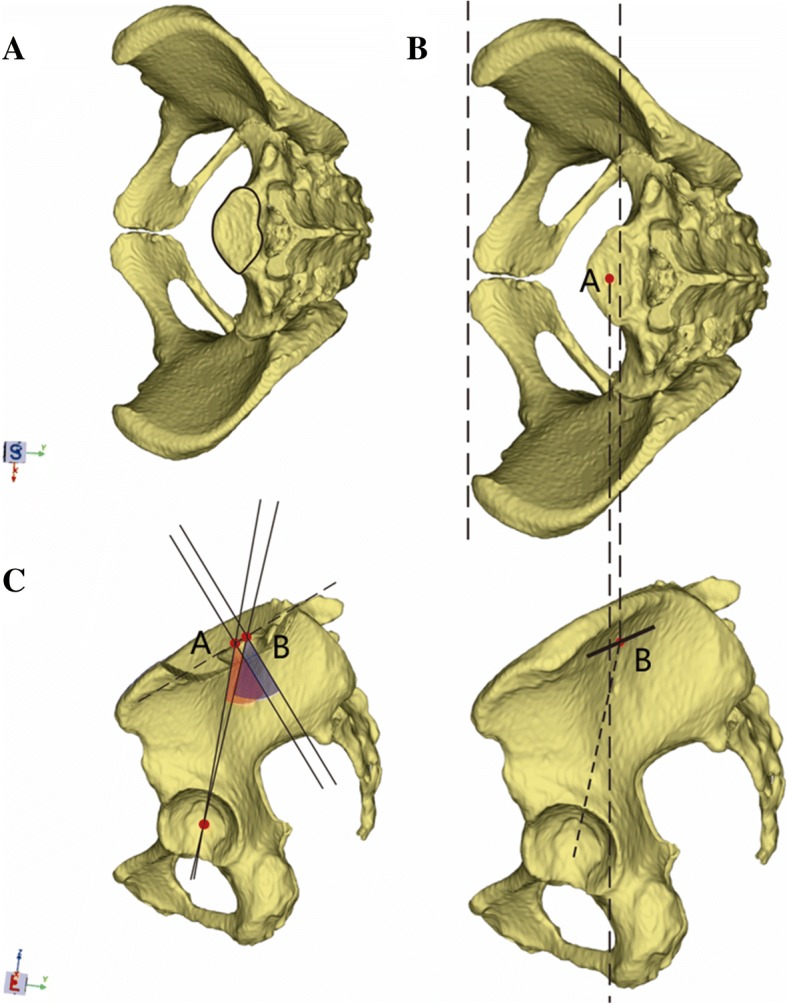
a–c The schematic shows the type 2 sacral endplate and the measurement of pelvic incidence based on CT and plain film X-ray methods. a Type 2 sacral endplate with a concave side posteriorly. b Point A is obtained as the midpoint of the segment crossing the mid-sagittal plane of the endplate via CT. Point B is obtained as the midpoint of the projection line via plain film X-ray method. c Point B is behind point A. Thus, the pelvic incidence value measured on CT is smaller than that measured via plain film X-ray