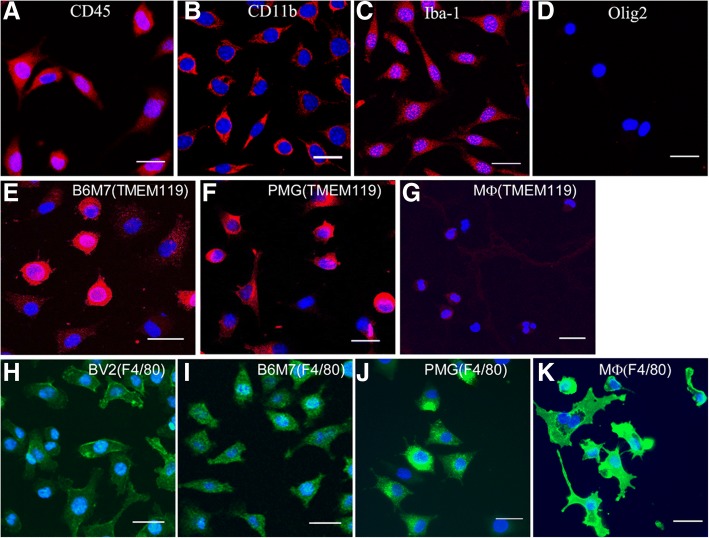
Fig. 1.
Immunocytochemistry of B6M7 cells. a-d B6M7 cells were stained for CD45 (a), CD11b (b), Iba1 (c), and Olig2 (d). e-g TMEM119 expression (red) in B6M7, primary microglia (PMG) and peritoneal macrophages (MФ). (h-k) F4/80 expression (green) in BV2, B6M7, PMG and peritoneal macrophages. Cell nuclei were stained with DAPI. All samples were examined by confocal microscopy. Scale bar = 25 μm

