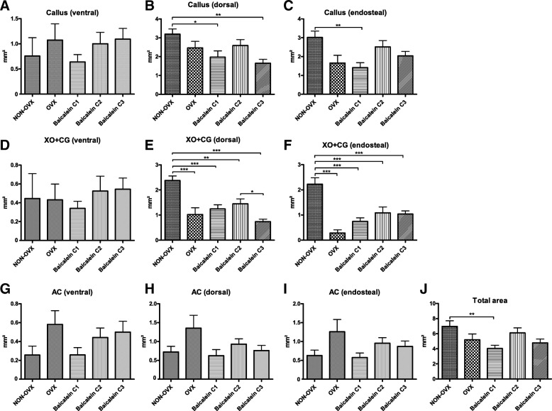
Fig. 6.
Histological analyses of fluorochrome-labelled sections of tibiae. The callus area was measured in ventral (a), dorsal (b) and endosteal aspects (c). Early stage of healing was measured in ventral (d), dorsal (e) and endosteal (f) aspects by XO + CG labelling and the late stage of healing by AC labelling (g-i). Finally, the total callus area was calculated (j). ***p ≤ 0.001, **p ≤ 0.01, *p ≤ 0.05 (NON-OVX n = 11, OVX n = 9, C1 n = 14, C2 n = 12, C3 n = 18)

