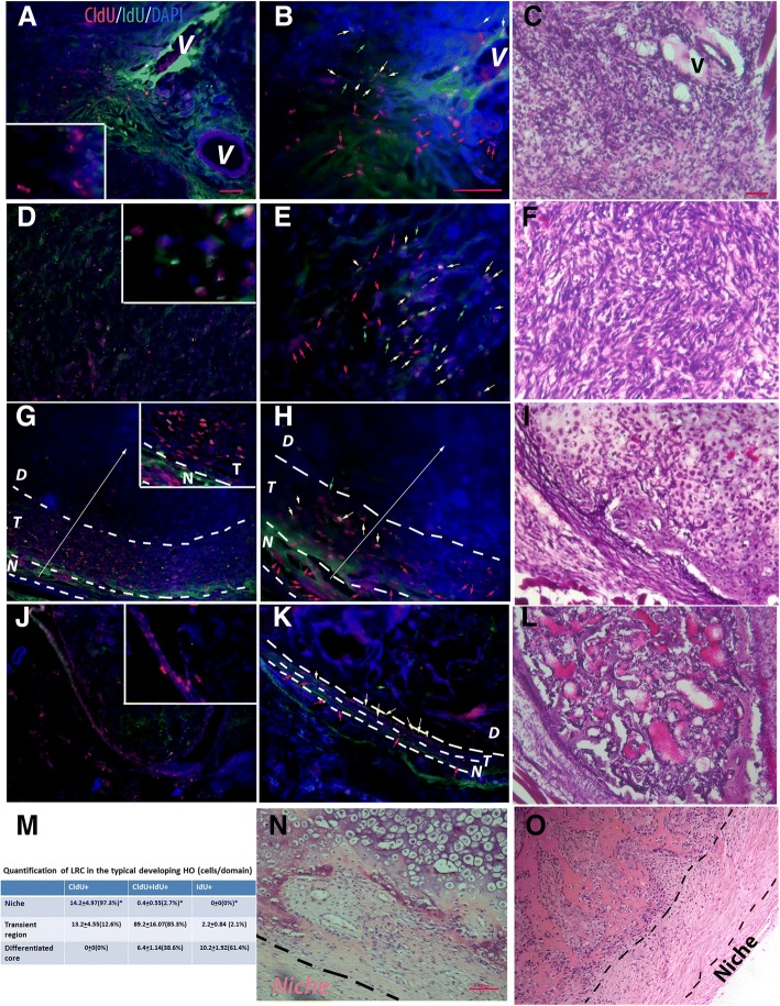
Fig. 1.
The patterns of LRCs in different stages of HO. a–c In the early inflammatory stage (< 3 days after injury) (a low power, b high power, and c H&E staining of the adjacent section), typical images showed many CldU+/IdU− cells (quiescent stem cells, red arrows), some CldU+/IdU+ (active stem cells, yellow arrows), but very few CldU−/IdU+ (transient amplifying cells, green arrows). Inserted box is a partial enlarged target area of panel a that highlights the staining pattern at this time point; note that no recognized regional pattern can be identified. d–f In the fibroproliferative stage (3–6 days after injury) (d low power, e high power, and f H&E staining of the adjacent section), typical images showed few CldU+/IdU− cells (red arrows), many CldU+/IdU+ (yellow arrows), and some CldU−/IdU+ (green arrows). Inserted box is a partial enlarged target area of panel d that highlights the staining pattern at this time point; note that no recognized regional pattern can be identified. g–i In developing HO (> 6 days after injury) (g low power, h high power, and i H&E staining of the adjacent section), the lesion could be roughly divided based on the consistent LRC pattern into three adjacent domains with a perceivable gradient from outside to the middle of the lesion (white arrows), i.e., the MSC niche (N), the transient domain (T), and the inner differentiated core (D). Inserted box is a partial enlarged target area of panel g that highlights the niche and the adjacent transient region; note the significant difference of staining pattern in these two different regions. (Also note the diffuse non-nuclei green staining is the background signal). Histologically, the MSC niche (N in g and h) was a very narrow strip. LRCs in this domain were almost exclusively CldU+/IdU− cells (red arrows). The transient amplifying domain (T in g and h), that was located between the MSC niche and the inner differentiated core, contained both CldU+/IdU+ (active stem cells, yellow arrows) and CldU−/IdU+ (transient amplifying cells, green arrows), with a perceivable gradient (white arrow), and the inner differentiated core (D in g and h) was composed of differentiated cells without significant thymidine analog label. j–l In mature HO (> 1 month after injury) (j low power, k high power, and l H&E staining of the adjacent section), the lesion could be similarly divided into three adjacent domains, except that the transient domain was often absent in the mature HO. Inserted box is a partial enlarged target area of panel j that highlights the narrow niche that contains almost exclusive the quiescent CldU+/IdU− cells and the adjacent region. m Quantification of different types of LRC in different domains of developing HO (2 weeks after injury). Average cell number/domain area in high-power image ± SD was shown (n = 5); percentages in parentheses represent the percentages of different subpopulations (CldU+, CldU+/IdU+, or IdU+) over the total LRC+ cells within each domain); statistics compared the percentages of positive cells in MSC domain vs. other domains, *Differ at p < 0.05 by ANOVA with Bonferroni post hoc testing. Note that in the proposed niche, 97.3% of total LRCs are CldU+ quiescent stem cells. In contract, in proposed transient region, most (89.2%) of LRCs are CldU+/IdU+ active stem cells, and in terminal differentiated core, the majority (61.4%) of LRCs are IdU+ transient amplifying cells. n, o A similar zonal pattern could be identified in human FOP (n) and aHO (o) samples, based on cell morphology (typical images of H&E staining shown). a, d, g, and j are on the same scale; b, e, h, and k are on the same scale; c, f, i, and l are on the same scale, bar = 50 μm. n and o are on the same scale, bar = 100 μm

