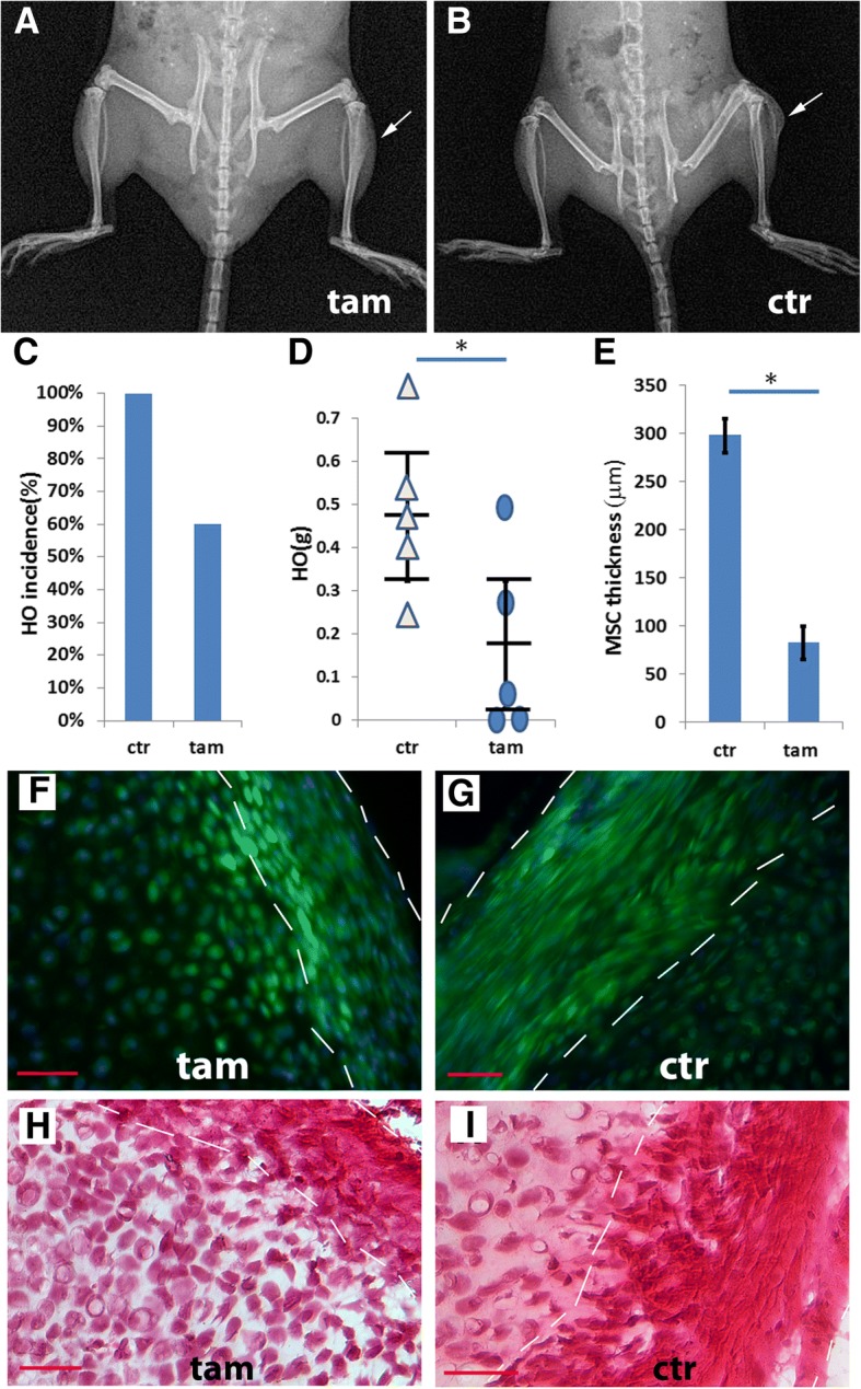
Fig. 3.

Conditional depletion of the Glast-creERT+ subpopulation inhibited injury-induced HO. a, b A typical X-ray image of TAM-treated (a) and control (b) Nse-BMP4;Glast-creERT;ROSA26-eGFP-DTA mice 4 weeks after injury. c HO incidence in control and TAM-treated groups. d Quantification of wet weight of HO in control and TAM treated groups, n = 5. Statistical analysis compared control (without TAM) vs. depleted (with TAM), using an unpaired t-test; *P < 0.05. Note that depletion of Glast-creERT+ cells inhibited but did not completely block HO. e Quantification of the thickness of MSC niche, n = 5, Statistical analysis compared control (without TAM) vs. depleted (with TAM), using an unpaired t-test; *P < 0.05. f Typical fluorescence images from TAM-treated (f) and control (g) Nse-BMP4;Glast-creERT;ROSA26-eGFP-DTA mice. Note that in the TAM-treated group (f), GFP− (recombined but not depleted) cells were only rarely found. h, i H&E staining of sections from TAM-treated (h) and control (i) Nse-BMP4;Glast-creERT;ROSA26-eGFP-DTA mice. Note that both the fluorescence images and the H&E staining suggest that the proposed MSC domain (within dashed white lines) was thinner in the TAM treated group. Bar = 50 μm
