
Fig. 6.
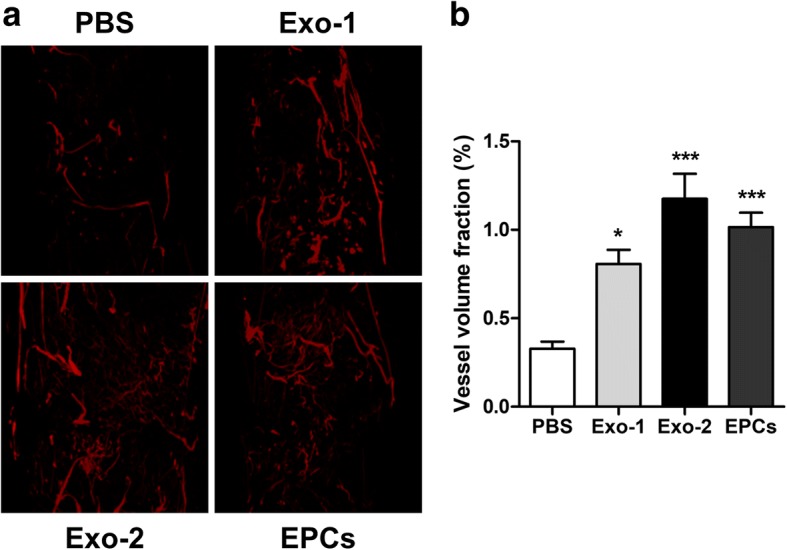
EPC-Exos enhanced the vessel density during distraction osteogenesis. a 3D reconstruction of the demineralized distraction regenerates perfused with Microfil at 4 weeks after consolidation. b Quantitative analysis of the vessel volume fractions within the distraction gaps from the four groups. *P < 0.05, ***P < 0.001 vs PBS group. EPCs endothelial progenitor cells, Exo exosomes secreted by EPCs, PBS phosphate-buffered saline