Figure 1.

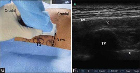
(a) High-frequency probe placed 3 cm lateral to T5 spinous process, (b) T – Trapezius, RM – Rhomboid major, ES – Erector spinae, TP – Transverse process, and P – Pleura

(a) High-frequency probe placed 3 cm lateral to T5 spinous process, (b) T – Trapezius, RM – Rhomboid major, ES – Erector spinae, TP – Transverse process, and P – Pleura