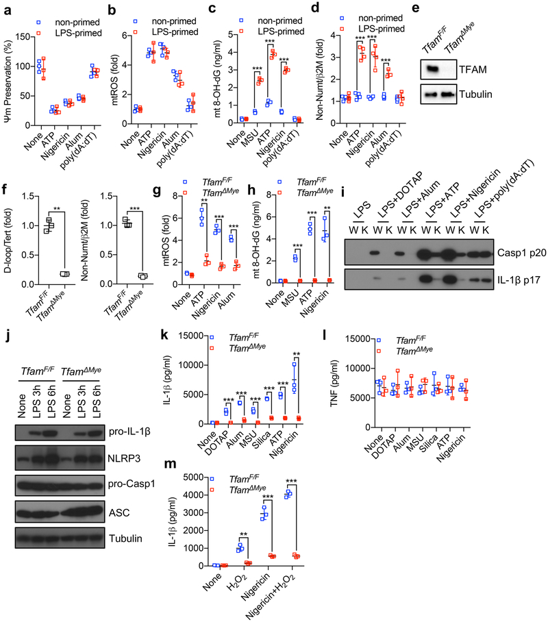
Extended Data Fig. 1 ~. TFAM is required for ox-mtDNA generation and NLRP3 inflammasome activation.
a, Inflammasome activator-induced changes in mitochondrial membrane potential (Ψm) in non-or LPS-primed wild-type BMDMs were measured by TMRM fluorescence. Data are mean ± s.d. (n = 3 biological replicates). b, Relative mtROS amounts were measured by MitoSOX fluorescence in non-or LPS-primed wild-type BMDMs after stimulation with different inflammasome activators. Data are mean ± s.d. (n = 3 biological replicates). c, Amounts of 8-OH-dG in mtDNA isolated from the mitochondrial fraction of non-or LPS-primed wild-type BMDMs that were treated with different inflammasome activators. Data are mean ± s.d. (n = 3 biological replicates). d, Cytosolic release of mtDNA, determined by qPCR with primers specific for mtDNA (non-NUMT) and nDNA (B2m), in non-or LPS-primed wild-type BMDMs after treatment with different inflammasome activators. Data are mean ± s.d. (n = 4 biological replicates). e, Immunoblot analysis of TFAM in Tfamff/f and TfamΔMye BMDMs. Results are typical of three independent experiments. f, Relative total mtDNA amounts in Tfamf/f and TfamΔMye BMDMs determined by qPCR with primers specific for mtDNA (D-loop, non-NUMT) and nDNA (Tert, B2m). Data are mean ± s.d. (n = 3 biological replicates). g, Relative mtROS amounts were measured by MitoSOX fluorescence in LPS-primed Tfamf/f and TfamΔMye BMDMs after stimulation with indicated NLRP3 activators. Data are mean ± s.d. (n = 3 biological replicates). h, Amounts of 8-OH-dG in mtDNA isolated from the mitochondrial fraction of LPS-primed Tfamff/f and TfamΔMye BMDMs that were stimulated with various NLRP3 activators. Data are mean ± s.d. (n = 3 biological replicates). i, Immunoblot analysis of Casp1 p20 and mature IL-1β (p17) in culture supernatants of Tfamf/f (W) and TfamΔMye (K) BMDMs that were stimulated with LPS plus different inflammasome activators. Results are typical of three separate experiments. j, Immunoblot analysis of pro-IL-1β, NLRP3, ASC and pro-Casp1 in the lysates of Tfamff/f and TfamΔMye BMDMs before and after LPS priming. Results are typical of three separate experiments. k, l, Amounts of IL-1β (k) and TNF (l) in culture supernatants of LPS-primed Tfamf/f and TfamΔMye BMDMs that were stimulated with various NLRP3 activators. Data are mean ± s.d. (n = 3 biological replicates). m, Amounts of IL-1β in culture supernatants of LPS-primed Tfamf/f and TfamΔMye BMDMs that were stimulated with H2O2 in the absence and presence of nigericin. Data are mean ± s.d. (n = 3 biological replicates). **P < 0.01; ***P < 0.001; two-sided unpaired t-test.

