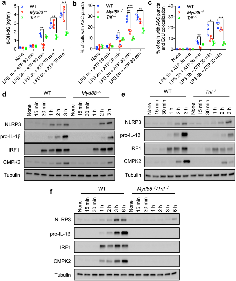
Extended Data Fig. 4 ~. MyD88 and TRIF mediate LPS-induced IRF1 and CMPK2 expression and NLRP3 inflammasome activation.
a, Amounts of 8-OH-dG in mtDNA isolated from mitochondrial fractions of wild-type, Myd88−/− and Trif−/− BMDMs that were primed with LPS for different durations followed by stimulation with ATP. Data are mean ± s.d. (n = 3 biological replicates per time point). b, c, Quantification of fluorescent microscopy images of ASC puncta (b) and ASC puncta positive for EdU (c) in wild-type, Myd88−/− and Trif−/− BMDMs that were primed with LPS for different durations, followed by ATP stimulation. Data are mean ± s.d. (n = 3 different microscopic fields per group; original magnification, ×40). d–f, Immunoblot analysis of NLRP3, pro-IL-1β, IRF1 and CMPK2 in wild-type, Myd88−/− and Trif−/− BMDMs before and after LPS stimulation. Data are typical of three separate experiments. *P < 0.05; **P < 0.01; ***P < 0.001; two-sided unpaired t-test.

