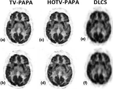
Figure 11.

Compressed sensing results for the patient brain phantom; (a), (b) reconstructed image from TV‐PAPA for small and large gap, respectively; (c), (d) reconstructed image from HOTV‐PAPA for small and large gap, respectively; (e), (f) reconstructed image from DL for small and large gap, respectively.
