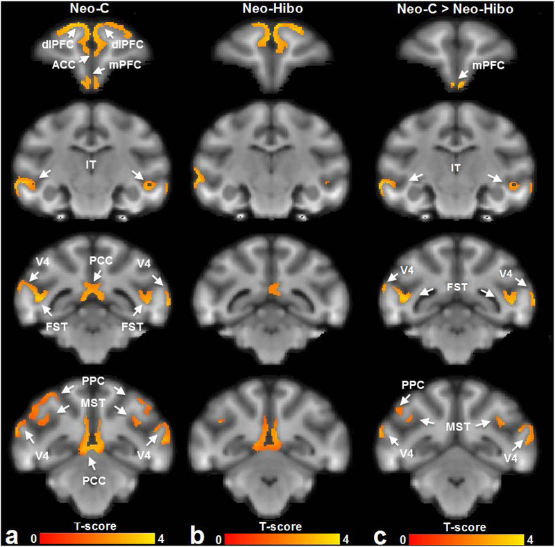
Figure 2.
The functional connectivity networks of dorsolateral prefrontal cortex derived from (a) animals with sham-operations Neo-C and (b) animals with Neo-Hibo lesions, by a one-sample t-test. A two-sample t-test (c) further showed that for the Neo-Hibo group functional connectivity decreased between dorsolateral prefrontal cortex (dlPFC) and medial prefontal cortex (mPFC), infero-temporal cortex (IT), fundus of superior temporal area (FST), posterior parietal cortex (PPC), medial superior temporal cortex (MST) and visual cortical area (V4). Other abbreviations: ACC: anterior cingulate cortex; PCC: posterior cingulate cortex. The brain template was generated with the T1-weighted images of Neo-C animals.

