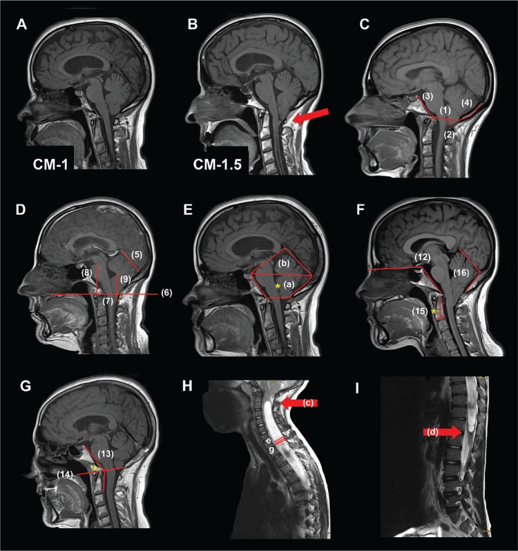
Figure 1. Measurements.
(A) Chiari malformation type 1 (CM-1): tonsillar descent (TD) ≥ 3 mm below the foramen magnum (FM) and the obex located above the level of the FM. (B) Chiari malformation type 1.5 (CM-1.5): TD ≥ 3 mm below the FM and obex located below the level of the FM. (C, D, E) Morphometric measurements made on mid-sagittal T1WI. Linear and planimetric parameters: (C-1) diameter FM; (C-2) cerebellar TD respect to the McRae line; (C-3) clivus length; (C-4) suboccipocium length; (D-5) tentorium length; (D-6) basal line (BL); (D-7) cerebellar TD respect to BL; (D-8) pons length; (D-9) fastigium length; (E-a) the osseous area of the posterior cranial fossa (PCF); (E-a+b) total PCF area. Angular measurements: (F) (F-12) basal angle; (G) (G-13) Wackenheim angle: (G-14*) basilar impression respect to the Chamberlain (F-15*) odontoid angle; (F-16) tentorium-occipital angle. Syringomyelia and spinal measurements: (H) (H-c, arrow) syringomyelia superior limit; (I) (I-d, arrow) syringomyelia inferior limit. Syringomyelia length = distance between superior and inferior limit. (H-e) Syringomyelia antero-posterior (AP) diameter; (H-f) spinal cord diameter: maximal diameter of the cord in the same slice that maximal diameter of the cavity in millimeters; (H-g) maximum spinal canal diameter. More specifications and descriptions of the morphometric measures are included in Text S1 in the supplemental material.

