
Figure 3.
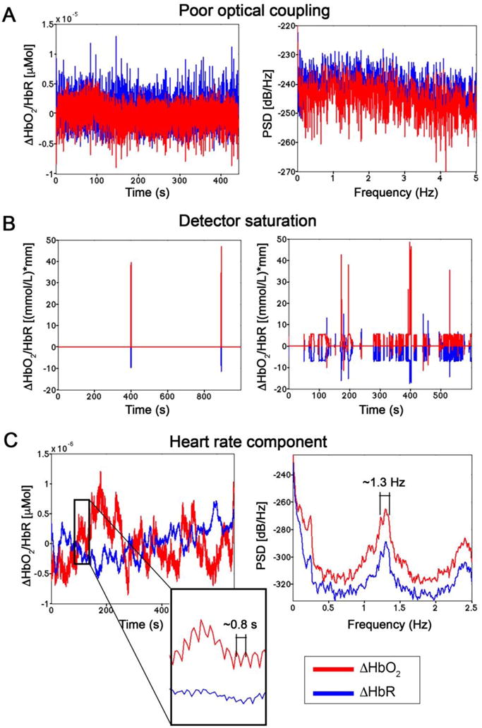
Example ΔHbO2 and ΔHbR in absence of a good coupling between the optodes and the head (A). This is reflected by the presence of only white noise, with a constant PSD. Data were in-house collected on the visual cortex using the Hitachi ETG-4000 during the presentation of a flashing checkerboard. In panel B, examples of channels corrupted by sunlight are shown, with consequent detector saturation. Data refer to the study by Pinti et al., 2015. The quality of fNIRS data can be assessed evaluating the presence of heart beat oscillations (C), visible both in the time- and in the frequency-domain. Data correspond to resting-state signals in-house recorded over the PFC using the Hitachi WOT-system.