. 2018 Dec 28;7:e38889. doi: 10.7554/eLife.38889

Figure 5. klf2b overexpression in endothelial cells can rescue the klf2 mutant cardiomyocyte extrusion phenotype.

(A–P) Endothelial- and myocardial-specific overexpression of klf2a or klf2b in klf2 WT and mutant hearts. Endothelial overexpression of klf2b (C–D, G–H). Myocardial overexpression of klf2a (K–L, O–P) or klf2b (I–J, M–N); maximum intensity projections of hearts in (A–D) and (I–L) are shown in (E–H) and (M–P), respectively. (Q–R’) Immunostaining of adult klf2 WT (Q–Q’) and rescued mutant (R–R’) heart sections for Caveolin1 to label epicardial cells and phalloidin for overall myocardial structure; magnified images of dashed boxes in (Q) and (R) are shown in (Q’) and (R’), respectively; arrows point to extruding cardiomyocytes; V: ventricle, At: atrium; scale bars: 50 µm (A–P), 300 µm (Q–R’).

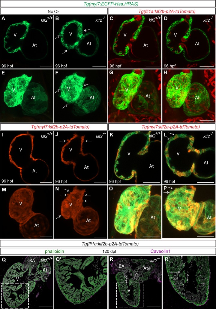
Figure 5.

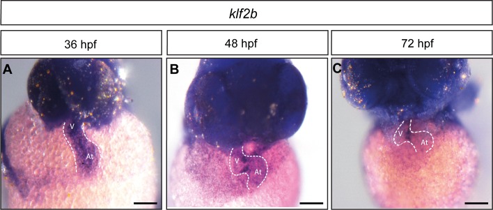
Figure 5—figure supplement 1. klf2b expression at early developmental stages .

Figure 5—figure supplement 1.

(A–C) Ventral views of in situ hybridization for klf2b expression at 36 (A), 48 (B) and 72 (C) hpf. V: ventricle, At: atrium; scale bars, 100 µm.
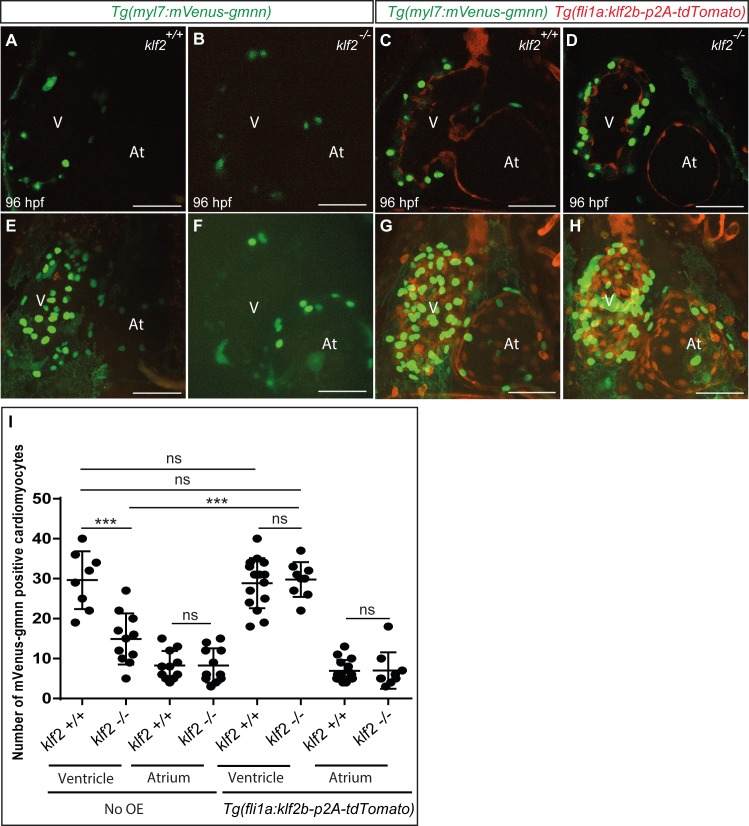
Figure 5—figure supplement 2. The cardiomyocyte proliferation defect in klf2 mutants is rescued by endothelial klf2b overexpression.

Figure 5—figure supplement 2.

(A–H) Confocal images (mid-sagittal sections) of 96 hpf klf2 WT (A, E, C and G) and mutant (B, F, D and H) hearts in the absence (A, B, E and F) or presence (C, D, G and H) of endothelial klf2b overexpression to examine cardiomyocyte proliferation. Maximum intensity projections of hearts in (A–D) are shown in (E-H). (I) Number of mVenus-gmnn positive ventricular and atrial cardiomyocytes in 96 hpf klf2 WT and mutant hearts in the absence or presence of fli1a:klf2b-p2a-tdTomato transgene; dots represent individual hearts; values represent means ±SEM; ***p≤0.001, ns (not significant), by Student’s t-test. V: ventricle, At: atrium; scale bars, 50 µm.
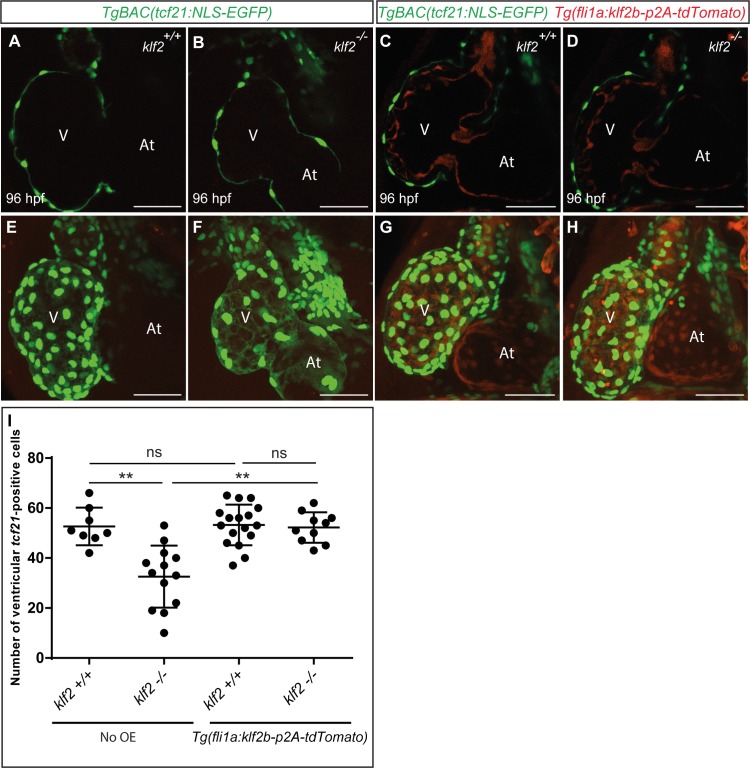
Figure 5—figure supplement 3. The epicardial defect in klf2 mutants is rescued by endothelial klf2b overexpression.

Figure 5—figure supplement 3.

(A–D) 2D confocal images (mid-sagittal sections) of 96 hpf klf2 WT (A, E, C and G) and mutant (B, F, D and H) hearts in the absence (A, B, E and F) or presence (C, D, G and H) of fli1a:klf2b-p2a-tdTomato transgene to examine cardiomyocyte proliferation. Maximum intensity projections of hearts in (A–D) are shown in (E-H). (I) Number of ventricular tcf21-GFP positive cells in 96 hpf animals plotted as a graph; dots represent individual hearts; values represent means ±SEM; **p≤0.01, ns (not significant), by Student’s t-test. V: ventricle, At: atrium; scale bars, 50 µm.
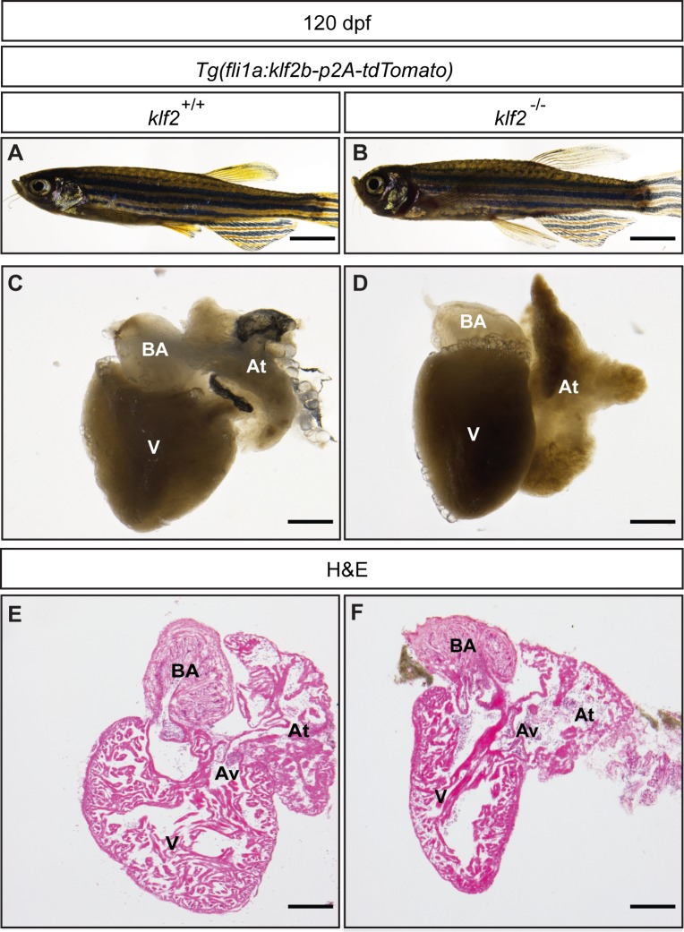
Figure 5—figure supplement 4. klf2 mutants rescued by endothelial klf2b overexpression can survive to adulthood.

Figure 5—figure supplement 4.

(A–B) Representative brightfield images of 120 dpf Tg(fli1a:klf2b-p2A-tdTomato); klf2 WT (A) and mutant (rescued by endothelial klf2b overexpression) (B) animals. (C–D) Adult Tg(fli1a:klf2b-p2A-tdTomato); klf2 WT (C) and rescued mutant (D) hearts from fish shown in (A and B), respectively. (E–F) Hematoxylin and eosin staining of adult klf2 WT (E) and rescued mutant (F) heart sections. V: ventricle, At: atrium, Av: atrioventricular canal; BA: bulbus arteriosus; scale bars: 5 mm (A–B), 200 µm (C–F).