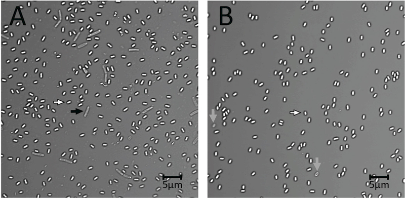
Figure 1A,B.

Photomicrograph of crude (A) and purified (B) B. subtilis spores. B. subtilis PS533 (Setlow and Setlow 1996) was sporulated on three 2xSG sporulation medium plates (Nicholson and Setlow 1990; Paidhungat et al. 2000) at 37°C. After 2 d spores were scraped from plates, suspended in ~ 30 ml cold water, sonicated for 3 min, centrifuged, the pellet suspended in 30 ml cold water and an aliquot photographed on an agarose-coated slide as described previously (Setlow et al. 2016; Li et al. 2017) either (A) immediately or (B) after spores were purified. For purification, the final suspension described above was sonicated for 3 min, centrifuged for 10 min at 17,000xg, suspended in 30 ml cold water with 3 min of sonication of the suspension followed by centrifugation and resuspension. The suspended spores at ~ 4°C were purified over 3 d by rounds of centrifugation, resuspension in cold water, sonication and centrifugation, ~ 3 rounds per d. In the last few d of this regimen, debris on the spore pellet surface was washed away by a gently spray of water. The scale bar in the figure is 5 microns, and white, grey and black arrows denote free dormant spores, germinated or lysed spores, and cells that did not sporulate, respectively. Note the debris in the background of panel A.
