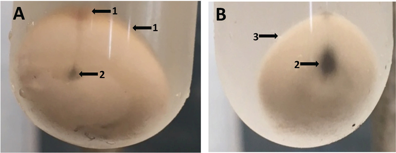
Fig. 2A,B.

Photographs of minimally (A) and fully (B) purified B. subtilis spore pellets. Three 2xSG medium sporulation plates as described in the legend of Fig. 1 were incubated at 37°C for 2 d, at which time the spores were scraped from the plates and suspended in ~ 30 ml of cold water. The cell forms in this suspension observed by bright field microscopy were ≥ 90% free spores as seen in Fig. 1A. The initial suspension was centrifuged for 10 min at 17,000xg, washed once by resuspension in cold water with 3 min of sonication of the suspension followed by centrifugation and resuspension. In tube A, the latter suspension was centrifuged in a 30 ml glass tube for 30 min at 6700xg and the pellet was photographed. The suspended spores were then further purified over a period of 3 d as described in the legend to Fig. 1. Finally, in B the purified spore suspension was centrifuged as described above for tube A and the pellet was photographed. Arrows labeled 1 in tube A indicate debris on the surface of the spore pellet, and the arrow labeled 2 in tube B shows that the surface of the extensively purified spore pellet is free from debris. The black material indicated by arrows labeled 3 in tubes A and B is small amounts of insoluble grit in sporulation media. This can be removed from spore suspensions, by allowing it to settle out from suspensions, as the grit settles out much faster that do spores.
