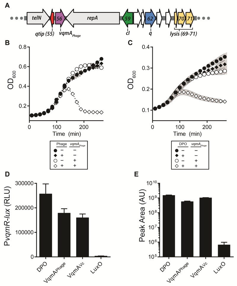
Figure 1: Phage VP882 encodes a homolog of vibrio VqmA that binds DPO and promotes host cell lysis.
(A) Organization of a region of the phage VP882 genome. Colors denote genes characterized here. telN and repA are conserved across linear plasmid-like phage. gp56 (purple) encodes the VqmAPhage QS receptor, gp55 (red) encodes the Qtip antirepressor, gp59 (green) encodes the cI repressor, gp62 (blue) encodes the Q antiterminator, and gp69-71 (yellow) encode the lysis genes. This color coding is used throughout this work. (B) Growth curves of V. parahaemolyticus lysogenized with phage VP882 (diamonds) or cured of phage VP882 (circles) harboring inducible vqmAPhage. As indicated, vqmAPhage expression was induced with 0.2% ara. (C) Growth curves of V. parahaemolyticus lysogenized with phage VP882 harboring inducible vqmAPhage in minimal medium lacking threonine. As indicated, vqmAPhage expression was induced with 0.2% ara and DPO was present at 10 μM. (D) Quantitation of DPO from purified proteins in a DPO-dependent E. coli reporter assay. The left bar shows the 10 μM synthetic DPO standard. RLU denotes relative light units. (E) Quantitation of DPO from the extracts in panel D by LC-MS. The left bar shows the 100 μM synthetic DPO standard. AU denotes arbitrary units. Data are represented as mean ± std with n=3 biological replicates (B, D), n=3 technical replicates (E), and n=3 biological and n=3 technical replicates (C). See also Figures S1 and S2.

