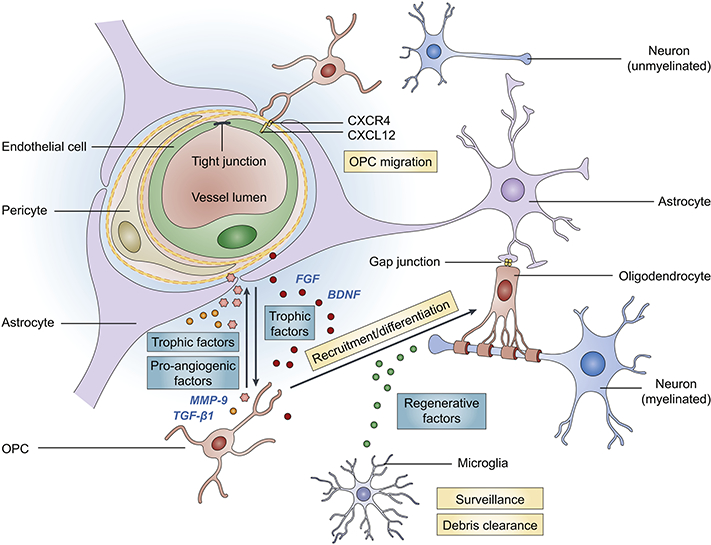
Fig. 1. Key cellular components of white matter and the oligovascular signaling.

The ‘oligovascular unit’ in the white matter consists of oligodendrocyte progenitor cells (OPCs), myelinating oligodendrocytes, endothelial cells, astrocytes, pericytes and microglia. These key cellular components are functionally coupled and influence each other under physiological and pathological conditions. Astrocytes and oligodendrocytes are coupled by gap junctions (Orthmann-Murphy et al., 2007; Robinson et al., 1993). Endothelial cells promote the survival, proliferation and differentiation of OPCs through secretion of trophic factors, e.g., brain-derived neurotrophic factor (BDNF) and fibroblast growth factor (FGF) (Arai and Lo, 2009b). On the other hand, OPCs support the survival of endothelial cells and alter the permeability of BBB via various trophic factors and pro-angiogenic factors, such as transforming growth factor (TGF)-ßl and matrix metallopeptidase (MMP)-9 (Seo et al., 2014; Seo et al., 2013). The migration of OPCs along vasculature and OPC-endothelium interaction coordinated by CXCR4-CXCL12 signaling is essential for normal brain development (Tsai et al., 2016). Pericytes also critically regulate OPC differentiation/maturation and white matter homeostasis (Maki et al., 2018). Pericyte deficiency disrupts white matter microcirculation and facilitates the deposition of toxic blood-derived substances, leading to a loss of myelin, axons and oligodendrocytes (Montagne et al., 2018). Microglia, the resident immune cells in the brain, actively patrol the white matter and produce regenerative factors that support OPC differentiation. Microglia also play a pivotal role in clearing myelin debris and facilitating remyelination after brain injury.
